Review
. 2015 Nov;23(11):1835-42.
doi: 10.1016/j.joca.2014.12.016.

Inflammation and intracellular metabolism: new targets in OA

Affiliations
Review

Inflammation and intracellular metabolism: new targets in OA

R Liu-Bryan. Osteoarthritis Cartilage. 2015 Nov.

Abstract

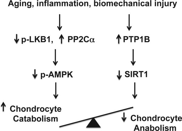
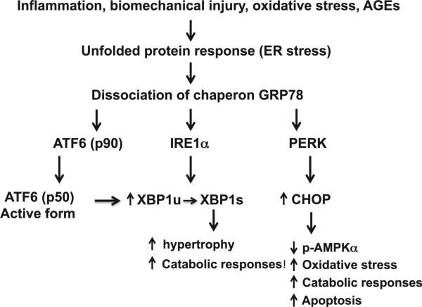
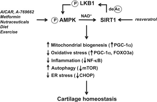
Articular cartilage degeneration is hallmark of osteoarthritis (OA). Low-grade chronic inflammation in the joint can promote OA progression. Emerging evidence indicates that bioenergy sensors couple metabolism with inflammation to switch physiological and clinical phenotypes. Changes in cellular bioenergy metabolism can reprogram inflammatory responses, and inflammation can disturb cellular energy balance and increase cell stress. AMP-activated protein kinase (AMPK) and sirtuin 1 (SIRT1) are two critical bioenergy sensors that regulate energy balance at both cellular and whole-body levels. Dysregulation of AMPK and SIRT1 has been implicated in diverse human diseases and aging. This review reveals recent findings on the role of AMPK and SIRT1 in joint tissue homeostasis and OA, with a focus on how AMPK and SIRT1 in articular chondrocytes modulate intracellular energy metabolism during stress responses (e.g., inflammatory responses) and how these changes dictate specific effector functions, and discusses translational significance of AMPK and SIRT1 as new therapeutic targets for OA.

Keywords: AMPK; Cartilage homeostasis; Energy metabolism; Inflammation; SIRT1.


Figures

Fig. 1
Fig. 1
Impaired chondrocyte AMPK and SIRT1 activity disrupts cartilage matrix homeostasis. Aging, inflammation and biomechanical injury in the joint can decrease phosphorylation of AMPKα Thr172 (indicating AMPK activity) and expression of SIRT1 in articular chondrocytes, likely through down-regulation of activity of the major upstream AMPK kinase LKB1 and up-regulation of protein phosphatase PP2Cα, and PTB1B. As a result, the balance between chondrocyte catabolic and anabolic function is disrupted. Overactive chondrocyte catabolic responses ultimately lead to cartilage matrix degradation.
Fig. 2
Fig. 2
The UPR in articular chondrocytes in OA. ER stress in OA chondrocytes caused by certain inflammatory mediators, biomechanical injury, nitric oxide and AGEs leads to activation of the UPR signaling cascades triggered by dissociation of the chaperon GRP78 from the ER transmembrane proteins PERK, IRE1α, and ATF6. The active XBP1 spliced form (XBP1s), generated by IRE1a through alternatively splicing mRNA of unspliced XBP1 (XBP1u), promotes chondrocyte hypertrophy and increases catabolic responses. The cleaved active form of ATF6 (p50) can induce XBP1 expression through direct binding to XBP1 promoter. Increased CHOP expression potentiates IL-1β to decrease phosphorylation of AMPKα and induce catabolic responses, superoxide generation and apoptosis.
Fig. 3
Fig. 3
AMPK and SIRT1 activity in regulation of cartilage homeostasis by increasing chondrocyte stress resistance. Pharmacological activators (e.g., AICAR, A-769662, and metformin), possibly some natural plant products (nutraceuticals), caloric restriction and exercise, activate AMPK in articular chondrocytes. As a result, intracellular NAD+ level is increased, leading to activation of SIRT1. Resveratrol, a natural plant product, also activates SIRT1. In turn, SIRT1 activates LKB1, the major upstream kinase of AMPK, through deacetylation of LKB1. Activation of AMPK and SIRT1 coordinates several housekeeping mechanisms to increase chondrocyte stress resistance by increasing mitochondrial biogenesis and function via PGC-1α; inhibiting oxidative stress and via PGC-1α and FOXO3a; attenuating inflammatory responses via inhibition of NF-κB activation, alleviating ER stress via limiting excessive CHOP; and promoting autophagy via suppression of mTOR, ultimately leading to cartilage homeostasis.

References

    1. Loeser RF, Goldring SR, Scanzello CR, Goldring MB. Osteoarthritis: a disease of the joint as an organ. Arthritis Rheum. 2012;64:1697–707. - PMC - PubMed
    1. Sellam J, Berenbaum F. The role of synovitis in pathophysiology and clinical symptoms of osteoarthritis. Nat Rev Rheumatol. 2010;6:625–35. - PubMed
    1. Scanzello CR, Goldring SR. The role of synovitis in osteoarthritis pathogenesis. Bone. 2012;51:249–57. - PMC - PubMed
    1. Goldring MB, Goldring SR. Articular cartilage and subchondral bone in the pathogenesis of osteoarthritis. Ann N Y Acad Sci. 2010;1192:230–7. - PubMed
    1. Liu-Bryan R, Terkeltaub R. The growing array of innate in-flammatory ignition switches in osteoarthritis. Arthritis Rheum. 2012;64:2055–8. - PMC - PubMed

Publication types