. 2015 Nov 2:5:15972.
doi: 10.1038/srep15972.

Long Non Coding RNA MALAT1 Promotes Tumor Growth and Metastasis by inducing Epithelial-Mesenchymal Transition in Oral Squamous Cell Carcinoma

Affiliations

Long Non Coding RNA MALAT1 Promotes Tumor Growth and Metastasis by inducing Epithelial-Mesenchymal Transition in Oral Squamous Cell Carcinoma

Xuan Zhou et al. Sci Rep. .

Abstract

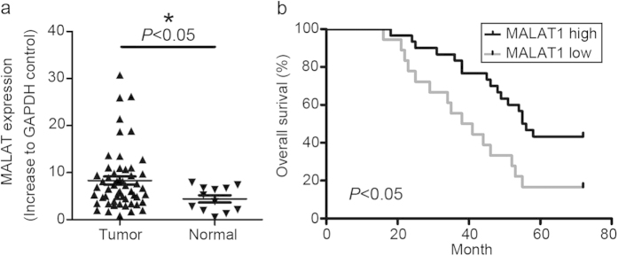
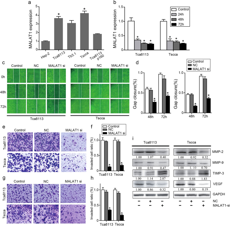
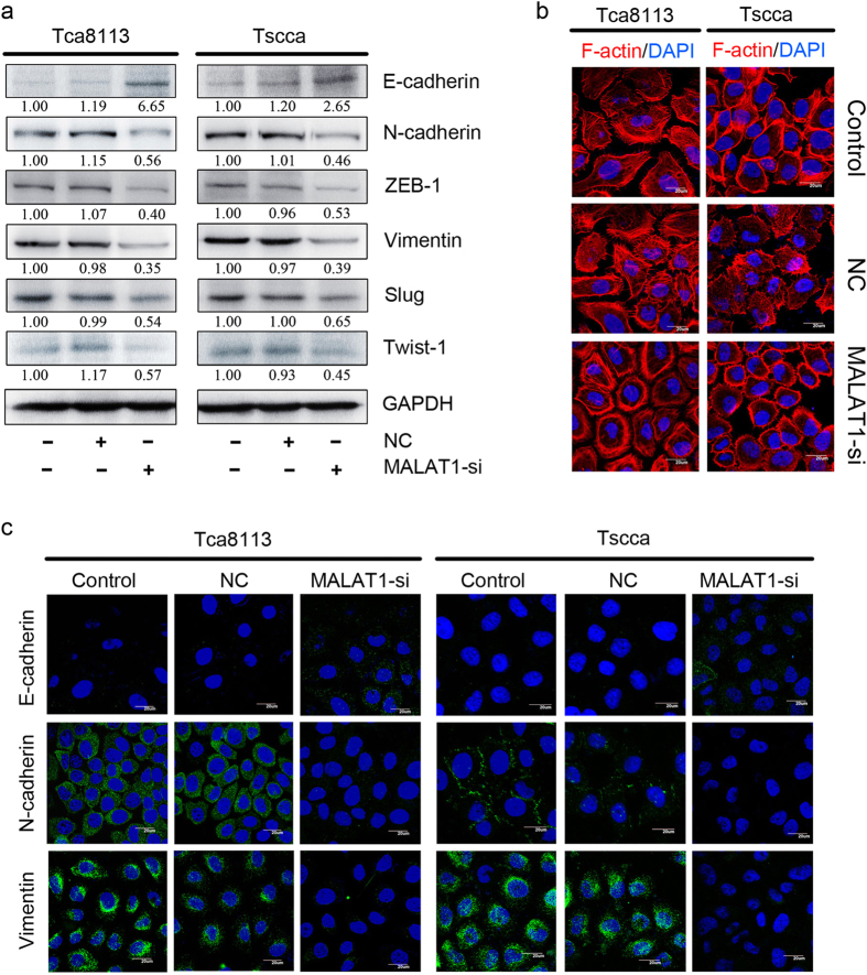
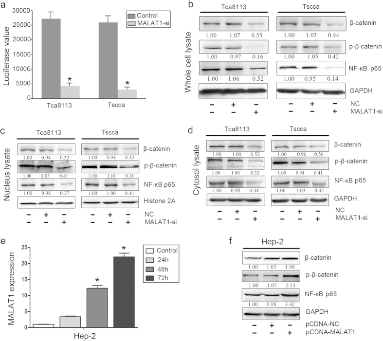
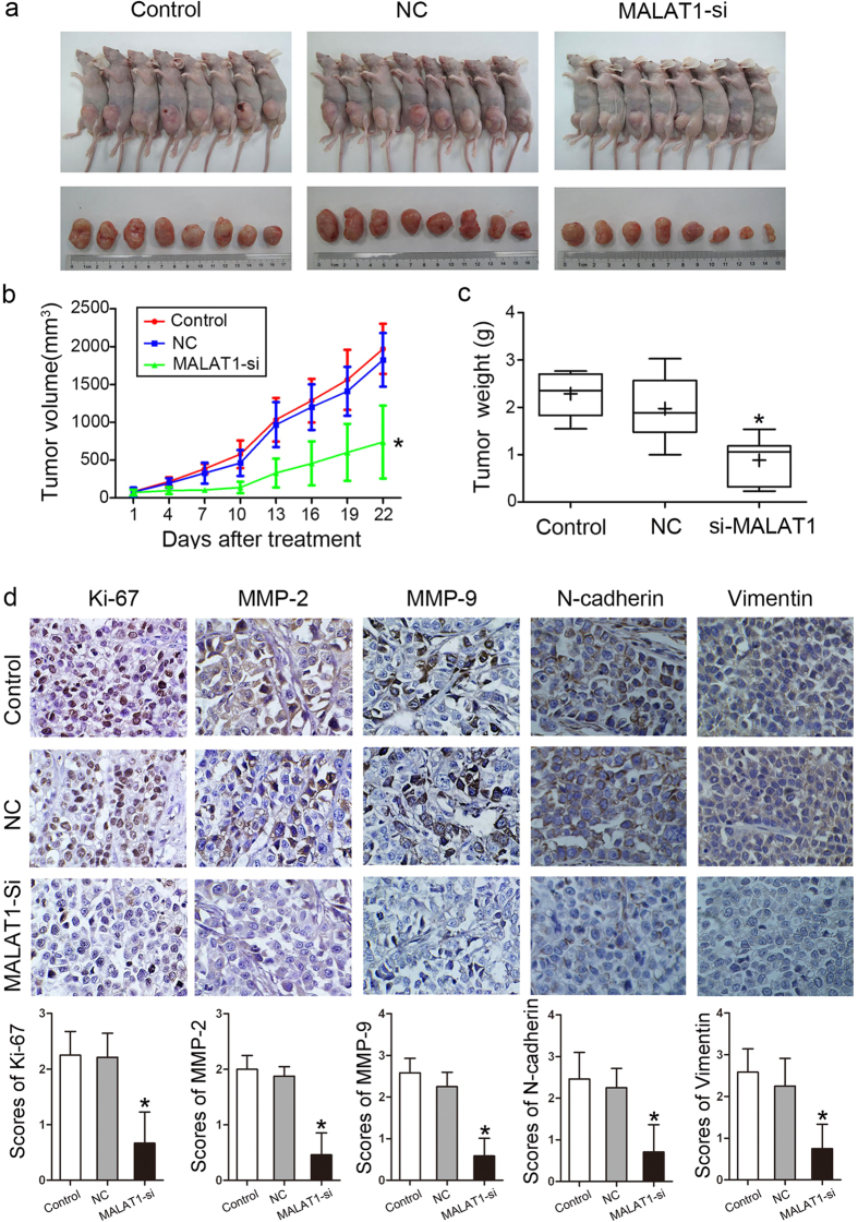
The prognosis of advanced oral squamous cell carcinoma (OSCC) patients remains dismal, and a better understanding of the underlying mechanisms is critical for identifying effective targets with therapeutic potential to improve the survival of patients with OSCC. This study aims to clarify the clinical and biological significance of metastasis-associated long non-coding RNA, metastasis-associated lung adenocarcinoma transcript 1 (MALAT1) in OSCC. We found that MALAT1 is overexpressed in OSCC tissues compared to normal oral mucosa by real-time PCR. MALAT1 served as a new prognostic factor in OSCC patients. When knockdown by small interfering RNA (siRNA) in OSCC cell lines TSCCA and Tca8113, MALAT1 was shown to be required for maintaining epithelial-mesenchymal transition (EMT) mediated cell migration and invasion. Western blot and immunofluorescence staining showed that MALAT1 knockdown significantly suppressed N-cadherin and Vimentin expression but induced E-cadherin expression in vitro. Meanwhile, both nucleus and cytoplasm levels of β-catenin and NF-κB were attenuated, while elevated MALAT1 level triggered the expression of β-catenin and NF-κB. More importantly, targeting MALAT1 inhibited TSCCA cell-induced xenograft tumor growth in vivo. Therefore, these findings provide mechanistic insight into the role of MALAT1 in regulating OSCC metastasis, suggesting that MALAT1 is an important prognostic factor and therapeutic target for OSCC.


Figures

Figure 1
Figure 1. Expression of MALAT1 in human oral squamous cell carcinoma.
(a) MALAT1 was overexpressed in 54 OSCC samples (P < 0.05). (b) Kaplan–Meier survival curve analysis indicated that OSCC patients with lower MALAT1 expression had prolonged survival time compared with patients with high levels of MALAT1 (log-rank test, P < 0.05).
Figure 2
Figure 2. Inhibition of MALAT1 attenuated OSCC invasion and migration in vitro.
(a) Tscca and Tca8113 cells displayed highest expression levels of MALAT1 among the HNSCC cell lines. (b) MALAT1 siRNA transfection inhibited effectively MALAT1 expression in Tscca and Tca8113 cells (P < 0.05). (c,d) MALAT1 siRNA significantly inhibited Tscca and Tca8113 cell migration determined by wound healing assay (P < 0.05). (e,f) MALAT1 siRNA significantly inhibited Tscca and Tca8113 cell migration and invasion determined by transwell assays with matrigel (P < 0.05). (g,h) MALAT1 siRNA significantly inhibited Tscca and Tca8113 cell migration determined by transwell assays without matrigel (P < 0.05). (i) MALAT1 siRNA significantly inhibited the expression of MMP2/9, and VEGF, but increased the expression of TIMP-3 in Tscca and Tca8113 cells determined by western blots analysis (P < 0.05). The gels were crapped, but those gels were run under the same experimental condition.
Figure 3
Figure 3. Inhibition of MALAT1 suppressed EMT in OSCC in vitro.
(a) MALAT1 siRNA significantly increased the expression of E-cadherin and inhibited the expression of N-cadherin, ZEB1, Vimentin, Slug, and Twist-1 in Tscca and Tca8113 cells determined by western blots analysis(P < 0.05). The gels were crapped, but those gels were run under the same experimental condition. (b) F-actin staining showed a stress-fiber pattern in control or NC siRNA treated cells, whereas a cortical pattern in MALAT1 siRNA-treated cells by immunofluorescence staining. (c) MALAT1 siRNA significantly upregulated E-cadherin and downregulated N-cadherin and Vimentin in Tscca and Tca8113 cells determined by immunofluorescence staining (original magnification: 1000×).
Figure 4
Figure 4. Inhibition of MALAT1 reduced the activity and level of β-catenin and NF-κB in OSCC.
(a) A TOP flash reporter assay indicated that MALAT1 siRNA treatment inhibited β-catenin transcriptional activity in Tscca and Tca8113 cells (P < 0.05). (b) MALAT1 siRNA treatment inhibited β-catenin, p-β-catenin and NF-kBp65 expression in whole cell lysate of Tscca and Tca8113 cells (P < 0.05). (c) MALAT1 siRNA treatment inhibited β-catenin, p-β-catenin and NF-kBp65 expression in nucleus lysate of Tscca and Tca8113 cells (P < 0.05). (d) MALAT1 siRNA treatment inhibited β-catenin, p-β-catenin and NF-kBp65 expression in cytosol lysate of Tscca and Tca8113 cells (P < 0.05). (e) PCDNA3-MALAT1 vector transfection increased MALAT1 expression in Hep-2 cells with a relative low level of MALAT1 expression (P < 0.05). (f) PCDNA3-MALAT1 vector transfection increased the expression of β-catenin, p-β-catenin and NF-kBp65 in whole lysate of Hep-2 cells (P < 0.05). The gels were crapped, but those gels were run under the same experimental condition.
Figure 5
Figure 5. MALAT1 knockdown inhibited tumor growth in a xenograft model in vivo.
(a) The image of Tscca xenograft tumors treated with si-NC and si-MALAT1. (b) Tumor growth curve indicated that MALAT1 siRNA injection treatment inhibited significantly Tscca xenograft tumor growth (P < 0.05). (c) Average tumor weight of si-MALAT1 group was reduced compared to the control and si-NC groups (P < 0.05). (d) IHC staining showed the expression of Ki-67, MMP-2, MMP-9, N-cadherin and Vimentin were inhibited in the MALAT1 siRNA-treated Tscca xenograft tumors (original magnification: 200×).

References

    1. Warnakulasuriya S. Global epidemiology of oral and oropharyngeal cancer. Oral Oncol 45, 309–316 (2009). - PubMed
    1. Leemans C. R., Braakhuis B. J. & Brakenhoff R. H. The molecular biology of head and neck cancer. Nat Rev Cancer 11, 9–22 (2011). - PubMed
    1. Acloque H. et al. Epithelial-mesenchymal transitions: the importance of changing cell state in development and disease. J Clin Invest 119, 1438–1449 (2009). - PMC - PubMed
    1. Polyak K. & Weinberg R. A. Transitions between epithelial and mesenchymal states: acquisition of malignant and stem cell traits. Nat Rev Cancer 9, 265–273 (2009). - PubMed
    1. Wang Y. et al. Akt/Ezrin Tyr353/NF-kappaB pathway regulates EGF-induced EMT and metastasis in tongue squamous cell carcinoma. Br J Cancer 110, 695–705 (2014). - PMC - PubMed

Publication types