

Inherited defects of thyroxine-binding proteins
- PMID: 26522458
- PMCID: PMC4632647
- DOI: 10.1016/j.beem.2015.09.002
Inherited defects of thyroxine-binding proteins
Abstract
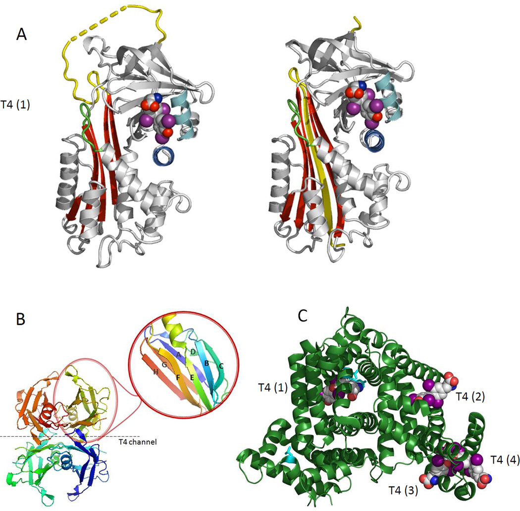
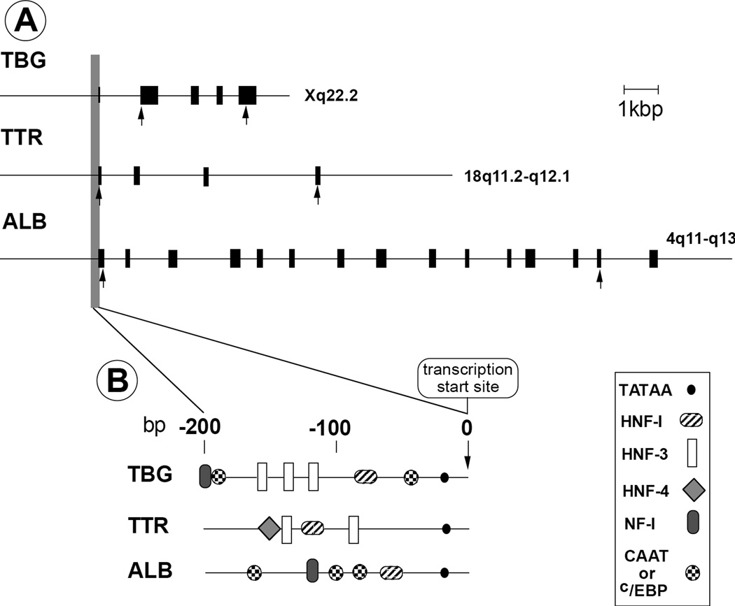
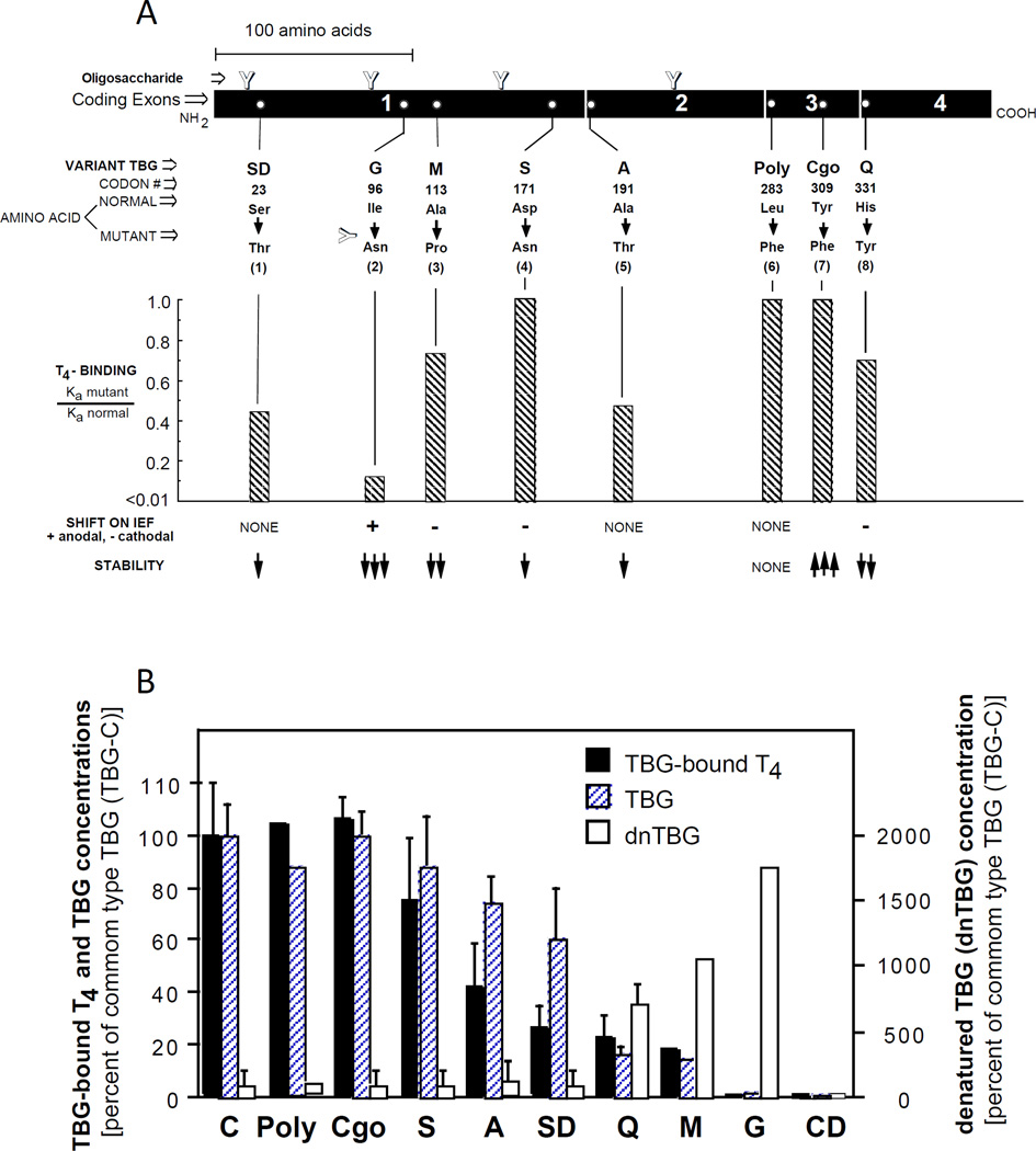
Thyroid hormones (TH) are bound to three major serum transport proteins, thyroxine-binding globulin (TBG), transthyretin (TTR) and human serum albumin (HSA). TBG has the strongest affinity for TH, whereas HSA is the most abundant protein in plasma. Individuals harboring genetic variations in TH transport proteins present with altered thyroid function tests, but are clinically euthyroid and do not require treatment. Clinical awareness and early recognition of these conditions are important to prevent unnecessary therapy with possible untoward effects. This review summarizes the gene, molecular structure and properties of these TH transport proteins and provides an overview of their inherited abnormalities, clinical presentation, genetic background and pathophysiologic mechanisms.
Keywords: TBG deficiency; familial dysalbuminemic hyperthyroxinemia; human serum albumin; mutations; thyroid hormone transport proteins; thyroxine-binding globulin; transthyretin.
Copyright © 2015 Elsevier Ltd. All rights reserved.
Figures
References
-
- Oppenheimer JH. Role of plasma proteins in the binding, distribution, and metabolism of the thyroid hormones. New England Journal of Medicine. 1968;278:1153–1162. - PubMed
-
- Mendel CM, Weisinger RA, Jones AL, Cavalieri RR. Thyroid hormone-binding proteins in plasma facilitate uniform distribution of thyroxine within tissues: A perfused rat liver study. Endocrinology. 1987;120:1742–1749. - PubMed
-
- Janssen OE, Golcher HMB, Grasberger H, et al. Characterization of thyroxine-binding globulin cleaved by human leukocyte elastase. Journal of Clinical Endocrinology and Metabolism. 2002;87:1217–1222. - PubMed
-
- Sunthornthepvarakul T, Likitmaskul S, Ngowngarmratana S, et al. Familial dysalbuminemic hypertriiodothyroninemia: a new dominantly inherited albumin defect. Journal of Clinical Endocrinology and Metabolism. 1998;83:1448–1454. - PubMed
-
- Refetoff S. Inherited thyroxine-binding globulin (TBG) abnormalities in man. Endocrine Reviews. 1989;10:275–293. - PubMed
Publication types
MeSH terms
Substances
Grants and funding
LinkOut - more resources
Full Text Sources
Other Literature Sources
Medical
Research Materials
Miscellaneous
