. 2016 Jul 7;35(27):3535-43.
doi: 10.1038/onc.2015.414. Epub 2015 Nov 2.

mTORC1 directly phosphorylates and activates ERα upon estrogen stimulation

Affiliations

mTORC1 directly phosphorylates and activates ERα upon estrogen stimulation

A Alayev et al. Oncogene. .

Abstract

Breast cancer is the leading cause of cancer-related deaths among women. Approximately 75% of breast cancers are estrogen receptor-α (ERα) positive, underscoring the dependence of cancer cells on estrogen for growth and survival. Patients treated with endocrine therapy often develop resistance, either de novo or acquired, which in some cases is caused by aberrations within the growth factor signaling pathways. The mechanistic target of rapamycin complex 1 (mTORC1) has emerged as a critical node in estrogenic signaling. We have previously shown that mTORC1 can phosphorylate and activate ERα on S167 via its effector-the 40S ribosomal S6 kinase 1 (S6K1). Presently, we have uncovered a direct link between mTORC1 and ERα. We found that ERα binds to regulatory-associated protein of mTOR (Raptor) and causes it to translocate to the nucleus upon estrogen stimulation. In addition, we identified mTOR as the kinase that phosphorylates ERα on S104/106 and activates transcription of ER target genes. Our findings show a direct link between mTORC1 and ERα, which further implicates mTORC1 signaling in the pathogenesis of ER-positive breast cancer and provides rationale for FDA-approved use of mTORC1 inhibitors in combination with endocrine agents for treatment of this disease.


Conflict of interest statement

Conflict of Interest: Authors declare no conflict of interest

Figures

Figure 1
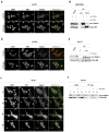
Figure 1. Raptor co-localizes with ERα in the nucleus upon estrogen stimulation
(a) MCF7 cells were serum-starved or stimulated with estrogen for 30 min. Immunofluorescence was performed as described in “Materials and Methods”. Scale bar represents 50μm. (b) BT474 cells were treated with estrogen as indicated and processed as described in (a). (c) MCF7 cells were treated with estrogen as indicated and processed as described in (a). (d) HEK293E cells were co-transfected with myc-raptor and FLAG-ER constructs as indicated. FLAG or myc was immunoprecipitated as described in “Materials and Methods” and proteins were detected by immunoblot. (e) MCF7 cells were co-transfected with myc-raptor and FLAG-ER and stimulated with estrogen for 30 min, as indicated. Raptor was immunoprecipitated and interaction with ERα is shown by immunoblot analysis. (f) MCF7 cells were co-transfected with myc-raptor and FLAG-ER, and nuclear (N) and cytoplasmic (C) fractionation was performed as described in “Materials and Methods”. Raptor was immunoprecipitated and interaction with ERα was detected by immunoblot.
Figure 2
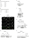
Figure 2. Tamoxifen disrupts ERα-Raptor interaction
(a) MCF7 cells were treated with tamoxifen for 1h and immunofluorescence was performed as described in “Materials and Methods”. Scale bar represents 50μm. (b) HEK293E cells were co-transfected with myc-raptor and FLAG-ER. Cells were treated with tamoxifen for 1h as indicated. ER was immunoprecipitated and interaction with raptor was detected by immunoblot.
Figure 3
Figure 3. mTOR forms a complex with Raptor and ERα
(a) HEK293E cells were co-transfected with AU-TOR, myc-raptor and FLAG-ER, and immunoprecipitated. (C is no antibody control). The indicated proteins were detected by immunoblot. (b) HEK293E cells were transfected with FLAG-ER, and after serum starvation, cells were stimulated with estrogen for 30 min. Immunofluorescence was performed as described in “Materials and Methods”. Scale bar represents 50μm.
Figure 4
Figure 4. mTORC1 interacts with ERα via the TOS motif and phosphorylates ERα on S104/106
(a) Alignment of TOS motif of ERα with TOS motifs of known mTOR targets. (b) Conservation of ERα TOS motifs. (c) HEK293E cells were transfected with WT or F/A ER. Immunofluorescence was performed as described in “Materials and Methods” with the indicated antibodies. (d) HEK293E cells were transfected with WT, F/A ER or S118A/S167A (SS/AA) alleles of ER and immunoprecipitated with FLAG. (C is no antibody control). The indicated proteins were detected by immunoblot. (e) HEK293E cells were transfected with WT or F/A ER. (C is empty vector). Lysates were generated as described in “Materials and Methods” and the indicated proteins were detected by immunblot. (f) Cells were treated with estrogen for 30 min as indicated. Immune complex kinase assay was performed as described in “Materials and Methods” using WT or F/A ER as substrate. (g) Cells were treated with estrogen for 30 min, immune complex kinase assay using WT or F/A ER as substrate was performed as described in “Materials and Methods” and the indicated proteins were detected by immunoblot. (h) HEK293E cells were transfected with WT or F/A ER and stimulated with estrogen for 30 min as indicated. Nuclear and cytoplasmic extraction was performed as described in “Materials and Methods” and indicated proteins were detected by immunoblot.
Figure 5
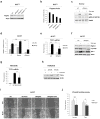
Figure 5. Raptor regulates ERα activity
(a) MCF7 cells were transfected with scrambled siRNA, or siRNAs against raptor alone or in combination and probed with the indicated antibodies. (b) Quantification of raptor normalized to actin from the western blot in (a) was performed using Odyssey Image Studio Version 4.0 and graphed using Excel. (c) MCF7 cells were transfected with either scrambled siRNA or siRNA against raptor. Cells were stimulated with estrogen for 30 min as indicated and probed with the indicated antibodies (d) MCF7 cells were transfected, stimulated with estrogen for 30 min as indicated and Luciferase reporter assay was performed as described in “Materials and Methods”. Data was plotted using Excel. * represents p<0.05 and ** represents p<0.01. (e) MCF7 cells were transfected with non-specific si or si against Raptor. RT-qPCR was performed as described in “Materials and Methods” and data was plotted using Excel. * represents p<0.05. ** represents p<0.001. n=3. (f) MCF7 cells were transfected as described in “Materials and methods”, stimulated with estrogen for 24 h as indicated and proteins levels were detected using immunoblot. (g) HEK293E cells were transfected with WT or F/A ER. (C is empty vector). RT-qPCR was performed as described in “Materials and Methods” and data were plotted using Excel. * represents p<0.05. n=3. (h) HEK293E cells were transfected as described in “Materials and Methods”, stimulated with estrogen for 24 h as indicated and protein levels were detected by immunoblot. (i) MCF7 cells were transfected with either scrambled siRNA or siRNA against raptor and treated with 4-hydroxy-tamoxifen (tam, 100nM) as indicated. Wound Healing Assay was performed as described in “Materials and Methods”. (j) Quantification of the Wound Healing Assay from (i) was plotted using Excel. * represents p<0.05. ** represents p<0.01.
Figure 6
Figure 6. Mechanism of ERα-raptor interaction
In the presence of estrogen, ERα binds to raptor via the TOS motif and recruits mTORC1 to the nucleus where mTOR phosphorylates ERα, leading to its activation and resulting in upregulation of estrogenic gene transcription.

References

    1. Couse JF, Korach KS. Estrogen receptor null mice: what have we learned and where will they lead us? Endocrine reviews. 1999;20(3):358–417. - PubMed
    1. Miller TW, Balko JM, Arteaga CL. Phosphatidylinositol 3-kinase and antiestrogen resistance in breast cancer. Journal of clinical oncology : official journal of the American Society of Clinical Oncology. 2011;29(33):4452–61. - PMC - PubMed
    1. Ellis M. Overcoming endocrine therapy resistance by signal transduction inhibition. The oncologist. 2004;9 (Suppl 3):20–6. - PubMed
    1. Miller WR, Larionov AA. Understanding the mechanisms of aromatase inhibitor resistance. Breast cancer research : BCR. 2012;14(1):201. - PMC - PubMed
    1. Maruani DM, Spiegel TN, Harris EN, Shachter AS, Unger HA, Herrero-Gonzalez S, et al. Estrogenic regulation of S6K1 expression creates a positive regulatory loop in control of breast cancer cell proliferation. Oncogene. 2012;31(49):5073–80. - PMC - PubMed

MeSH terms