. 2015 Nov 2:6:8728.
doi: 10.1038/ncomms9728.

Transmembrane protein sorting driven by membrane curvature

Affiliations

Transmembrane protein sorting driven by membrane curvature

H Strahl et al. Nat Commun. .

Abstract

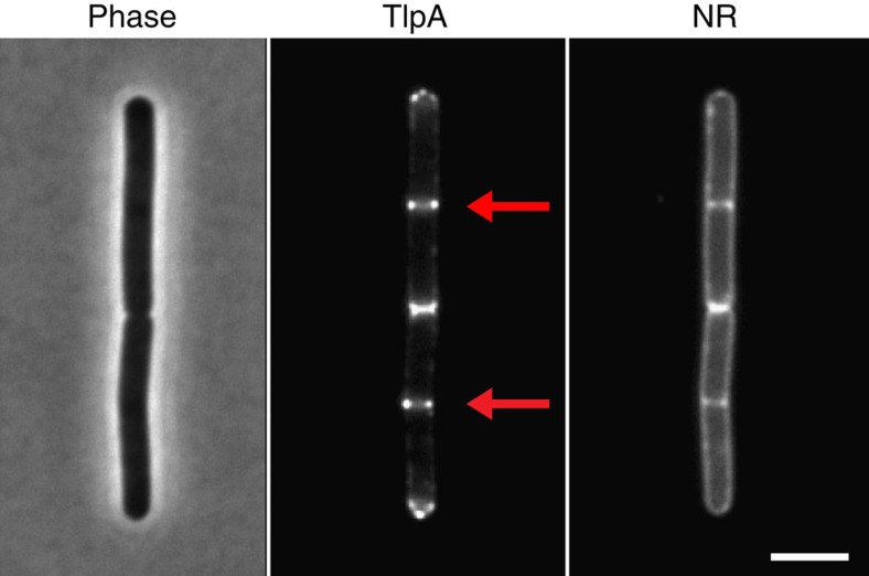
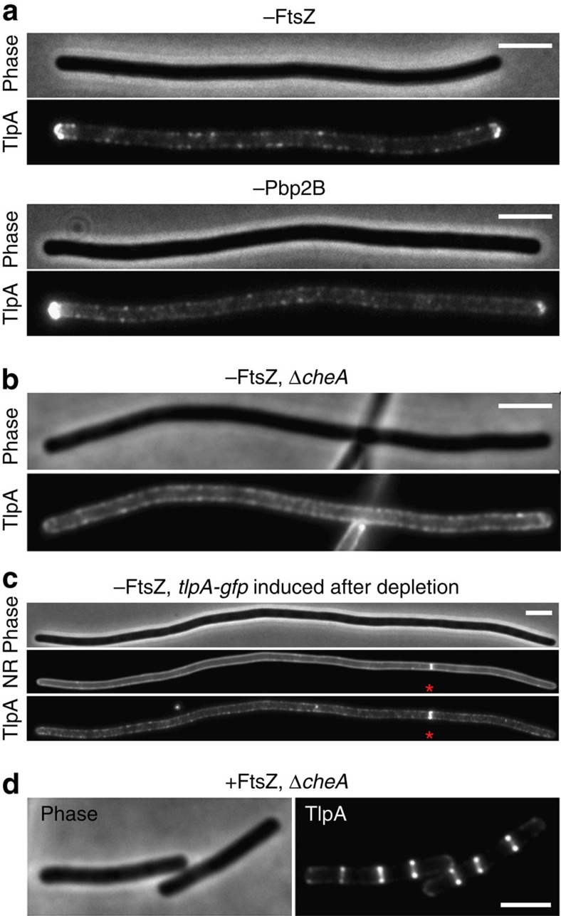
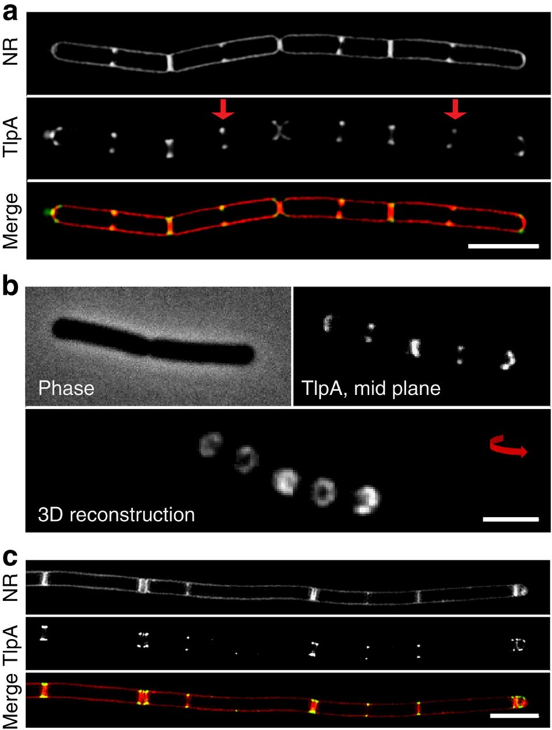
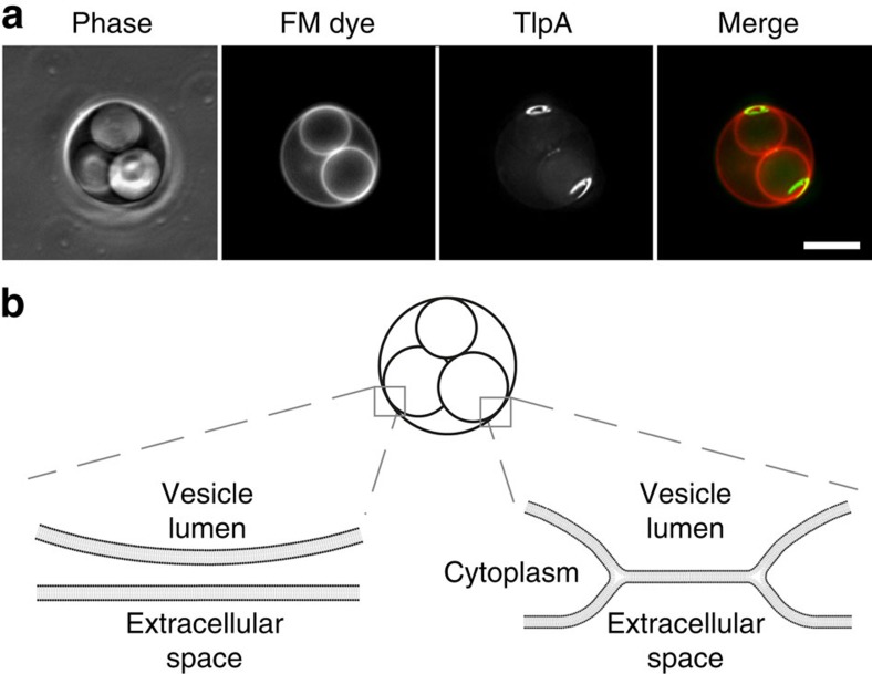
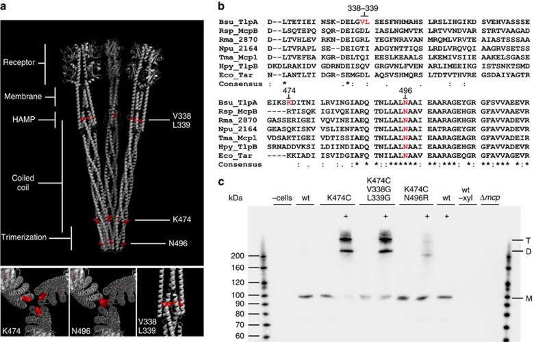
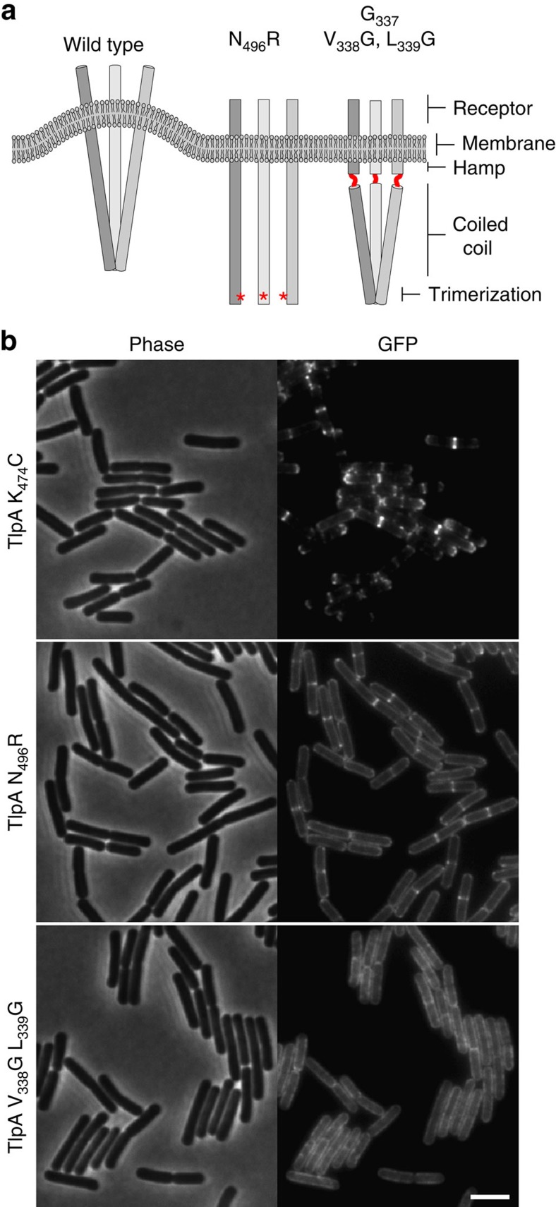
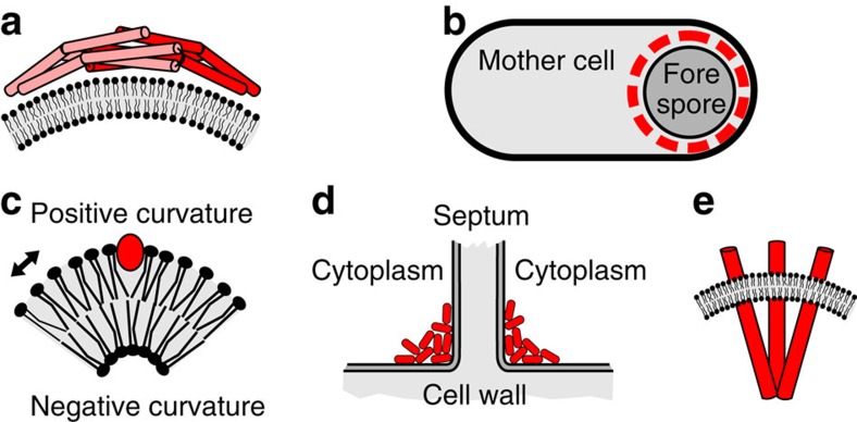
The intricate structure of prokaryotic and eukaryotic cells depends on the ability to target proteins to specific cellular locations. In most cases, we have a poor understanding of the underlying mechanisms. A typical example is the assembly of bacterial chemoreceptors at cell poles. Here we show that the classical chemoreceptor TlpA of Bacillus subtilis does not localize according to the consensus stochastic nucleation mechanism but accumulates at strongly curved membrane areas generated during cell division. This preference was confirmed by accumulation at non-septal curved membranes. Localization appears to be an intrinsic property of the protein complex and does not rely on chemoreceptor clustering, as was previously shown for Escherichia coli. By constructing specific amino-acid substitutions, we demonstrate that the preference for strongly curved membranes arises from the curved shape of chemoreceptor trimer of dimers. These findings demonstrate that the intrinsic shape of transmembrane proteins can determine their cellular localization.


Figures

Figure 1
Figure 1. TlpA localizes at cell poles and cell division sites.
Phase-contrast image of B. subtilis cells (left panel) expressing TlpA-GFP (middle panel), and stained with the fluorescent membrane probe Nile red (right panel). The active cell division sites are highlighted with an arrow. Strain used: B. subtilis HS48 (Pxyl-tlpA-gfp). Scale bar, 3 μm.
Figure 2
Figure 2. Localization of TlpA is cell division dependent.
(a) Midcell localization of TlpA-GFP is abolished in cells depleted for FtsZ (upper panel) and Pbp2B (lower panel). (b) Polar accumulation of TlpA-GFP is absent in cells depleted for FtsZ when cheA is deleted. (c) No recruitment to cell poles is observed when TlpA-GFP is induced after FtsZ depletion. However, a clear recruitment is observed to a single cell division site that is still present (indicated with an asterisk). (d) TlpA-GFP still localizes at cell division sites in a cheA deletion mutant. Strains used: (a) B. subtilis HS50 (Δmcp Pxyl-tlpA-gfp Pspac-ftsZ), B. subtilis HS51 (Δmcp Pxyl-tlpA-gfp Pspac-pbpB), (b/d) B. subtilis HS52 (ΔcheA Pxyl-tlpA-gfp Pspac-ftsZ) and (c) B. subtilis HS50 (Δmcp Pxyl-tlpA-gfp Pspac-ftsZ). Scale bar, 3 μm.
Figure 3
Figure 3. TlpA localizes to strongly curved septal membranes.
(a) Structured illumination microscopy (3D SIM) images of B. subtilis cells stained with Nile red (upper panel) and expressing TlpA-GFP (middle panel). Merged image shown in lower panel. (b) Phase-contrast image (upper left panel), confocal fluorescence image (upper right panel) and 3D reconstruction of optical sectioning of cells expressing TlpA-GFP (lower panel). (c) Localization of TlpA-GFP in the absence of DivIVA. 3D SIM images of Nile red membrane stained divIVA deletion mutant (upper panel) expressing TlpA-GFP (middle panel), and merged image (lower panel) are depicted. In the absence of DivIVA, B. subtilis cells frequently undergo multiple adjacent cell division events. Strains used: (a,b) B. subtilis HS49 (Δmcp Pxyl-tlpA-gfp) and (c) B. subtilis HS52 (Δmcp Pxyl-tlpA-gfp ΔdivIVA). Scale bar, 3 μm.
Figure 4
Figure 4. TlpA is recruited to non-septal curved membranes.
(a) The differential staining of internal vacuole-like structures with membrane dye FM 4–64 in B. subtilis L-forms indicates the presence of two distinct types of vacuoles within the cell. The membrane dye FM 4–64 is not able to cross biological membranes and therefore does not stain internal membrane structures, unless they remain connected to the cytoplasmic membrane. The connection with the cytoplasmic membrane (membrane hemifusion) generates membrane areas with a distinct curvature. TlpA-GFP is strongly recruited to these non-septal areas of high membrane curvature. Maximal intensity projection of a deconvolved optical sectioning is shown. Strain used: B. subtilis HS55 (Pxyl-tlpA-gfp L-form). Scale bar, 5 μm. (b) Schematic depiction of the interfaces between the vacuolar and cytoplasmic membranes. In the left, the membranes are fully separated resulting in a lack of FM 4–64 staining. In the right, the membranes remain attached via membrane hemifusion, allowing lateral diffusion of FM 4–64.
Figure 5
Figure 5. Complex formation analysis of TlpA mutants by in vivo crosslinking.
(a) Structural model of TlpA trimer of dimers with the position of the amino-acid substitutions highlighted in red, and close-up images of the same substitutions shown below. (b) ClustalW2 alignment of selected chemoreceptors representing examples from different bacterial phyla and classes. Bsu, B. subtilis (Firmicutes); Eco, E. coli (ϕ-Proteobacteria); Npu, Nostoc punctiforme (Cyanobacteria); Rsp, Rhodobacter sphaeroides (α-Proteobacteria); Rma, Rhodothermus marinus (Bacterioidetes); Tma, Thermotoga maritima (Thermatogae); Hpy, Helicobacter pylori (ɛ-Proteobacteria). The positions of the introduced amino-acid substitutions are highlighted in red. Note that the introduction of two glycine substitutions at positions 338 and 339 creates a stretch of three glycines. (c) Western blot of TlpA-GFP complexes carrying the N496R, and V338G L339G exchanges after in vivo crosslinking with TMEA. A cysteine residue was introduced at position 474, which allows tris(2-maleimidoethyl) amine (TMAE) crosslinking of neighbouring TlpA dimers if those are assembled into a trimer of dimers. Crosslinked samples are highlighted with ‘+'. As a control, Δmcp cells encoding wild-type TlpA-GFP with induction (wt), without induction (wt -xyl) and the parental strain (Δmcp) are shown. Monomeric TlpA-GFP (99 kDa), and crosslinked dimer and trimer bands are highlighted with M, D and T, respectively. Strains used: B. subtilis 168 (wild type), B. subtilis OI3545 (Δmcp), B. subtilis HS49 (Δmcp pxyl-tlpA-gfp), B. subtilis HS58 (Δmcp pxyl-tlpA(K474C)-gfp), B. subtilis HS59 (Δmcp pxyl-tlpA(K474C, N496R)-gfp) and B. subtilis HS60 (Δmcp pxyl-tlpA(K474C, V338G, L339G)-gfp).
Figure 6
Figure 6. Localization of TlpA depends on the shape of the protein complex.
(a) Schematic model of a TlpA trimer of dimers bending the cell membrane and the consequences of the different mutations (see main text for details). (b) Phase-contrast (left panel) and fluorescence (right panel) images of B. subtilis cells expressing TlpAK474C-GFP (cysteine substitution used for crosslinking), TlpAN496R-GFP (no trimerization) and TlpAV338G, L339G-GFP (increased flexibility). Strains used: B. subtilis HS58 (Δmcp Pxyl-tlpA(K474C)-gfp), B. subtilis HS59 (Δmcp Pxyl-tlpA(K474C, N496R)-gfp) and B. subtilis HS60 (Δmcp Pxyl-tlpA(K474C, V338G, L339G)-gfp). Scale bar, 3 μm.
Figure 7
Figure 7. Different mechanisms to localize at curved membranes.
(a) Schematic depiction of the BAR domain containing Arfaptin dimer using the ‘scaffolding' mechanism to generate and sense membrane curvature. (b) Sporulating B. subtilis cell with SpoVM (red) coated forespore. (c) Insertion of an amphipathic helix (red) at the positively curved lipid bilayer face. Arrow indicates increased spacing. (d) Accumulation of DivIVA (red) at negatively curved membranes at the base of a cell dividing septum. (e) Membrane curvature generated by the chemoreceptor trimer-of-dimers shape.

References

    1. Cobbold C., Monaco A. P., Sivaprasadarao A. & Ponnambalam S. Aberrant trafficking of transmembrane proteins in human disease. Trends Cell Biol. 13, 639–647 (2003). - PubMed
    1. Rudner D. Z. & Losick R. Protein subcellular localization in bacteria. Cold Spring Harb. Perspect. Biol. 2, a000307 (2010). - PMC - PubMed
    1. Kuwada N. J., Traxler B. & Wiggins P. A. Genome-scale quantitative characterization of bacterial protein localization dynamics throughout the cell cycle. Mol. Microbiol. 95, 64–79 (2015). - PMC - PubMed
    1. Shapiro L., McAdams H. H. & Losick R. Why and how bacteria localize proteins. Science 326, 1225–1228 (2009). - PMC - PubMed
    1. Govindarajan S., Elisha Y., Nevo-Dinur K. & Amster-Choder O. The general phosphotransferase system proteins localize to sites of strong negative curvature in bacterial cells. MBio 4, e00443–00413 (2013). - PMC - PubMed

Publication types

LinkOut - more resources