

Review
doi: 10.1155/2015/182872.
Epub 2015 Oct 7.
Nonmuscle Tissues Contribution to Cancer Cachexia
Affiliations
- PMID: 26523094
- PMCID: PMC4615210
- DOI: 10.1155/2015/182872
Item in Clipboard
Review
Nonmuscle Tissues Contribution to Cancer Cachexia
Mediators Inflamm.
2015.
Abstract
Cachexia is a syndrome associated with cancer, characterized by body weight loss, muscle and adipose tissue wasting, and inflammation, being often associated with anorexia. In spite of the fact that muscle tissue represents more than 40% of body weight and seems to be the main tissue involved in the wasting that occurs during cachexia, recent developments suggest that tissues/organs such as adipose (both brown and white), brain, liver, gut, and heart are directly involved in the cachectic process and may be responsible for muscle wasting. This suggests that cachexia is indeed a multiorgan syndrome. Bearing all this in mind, the aim of the present review is to examine the impact of nonmuscle tissues in cancer cachexia.
Figures
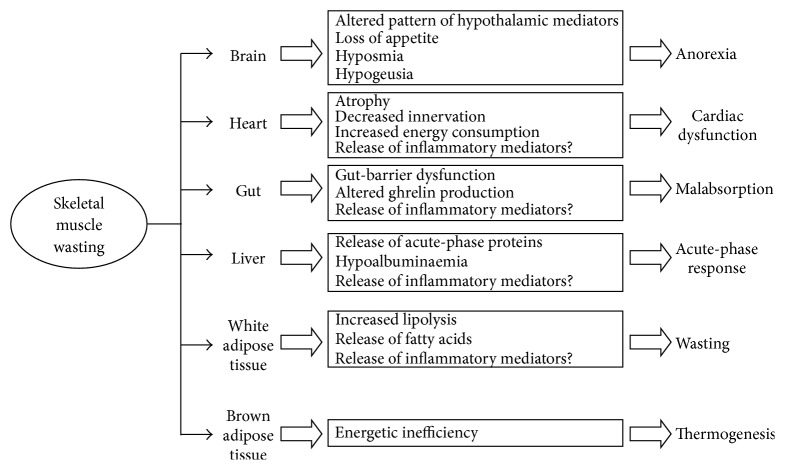
Interactions between different tissues/organs and skeletal muscle in the development of wasting associated with cancer cachexia. The events and metabolic alterations that take place in different tissues/organs during cancer cachexia may be related with the loss of muscle tissue. Indeed, muscle wasting may be influenced by the liver, inflammatory response, and by adipose tissues, particularly white fat. Brown adipose tissue could partially account for the energy inefficiency associated with hypermetabolism in the cancer patient. Brain, basically by modulation of appetite, also contributes to muscle wasting. The gut may be responsible for both malabsorption and changes in ghrelin production and, finally, the heart could also be a source of inflammatory mediators contributing to further muscle wasting.
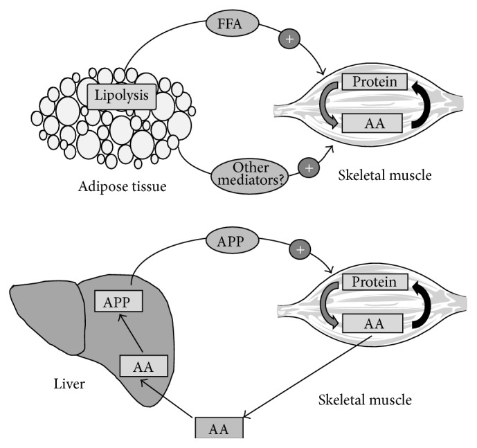
Examples of cross talk between adipose tissue/liver and skeletal muscle during cancer cachexia. Adipose tissue releases some factor(s), possibly fatty acids, that seem to be essential to activate muscle proteolysis. Indeed, recent evidences, using knockout deficient mice, suggest that blocking lipolysis in white fat results in an amelioration of muscle wasting. Similarly, liver acute-phase proteins (APP), such as serum amyloid A (SAA), could participate, alone or in synergy with cytokines, in activating muscle wasting by enhancing protein degradation.
References
-
- Warren S. The immediate cause of death in cancer. The American Journal of the Medical Sciences. 1932;184:610–613.
Publication types
MeSH terms
LinkOut - more resources
Full Text Sources
Other Literature Sources
