. 2016 Jan;17(1):87-94.
doi: 10.1038/ni.3310. Epub 2015 Nov 2.

Structural interplay between germline interactions and adaptive recognition determines the bandwidth of TCR-peptide-MHC cross-reactivity

Affiliations

Structural interplay between germline interactions and adaptive recognition determines the bandwidth of TCR-peptide-MHC cross-reactivity

Jarrett J Adams et al. Nat Immunol. 2016 Jan.

Abstract

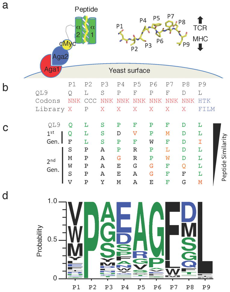
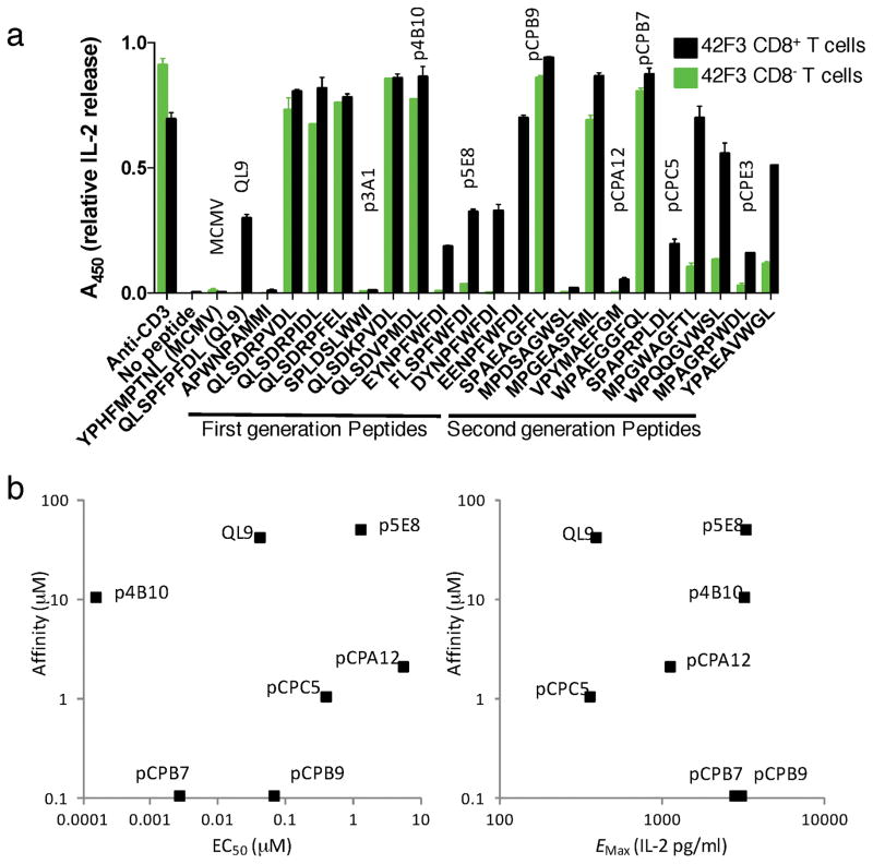
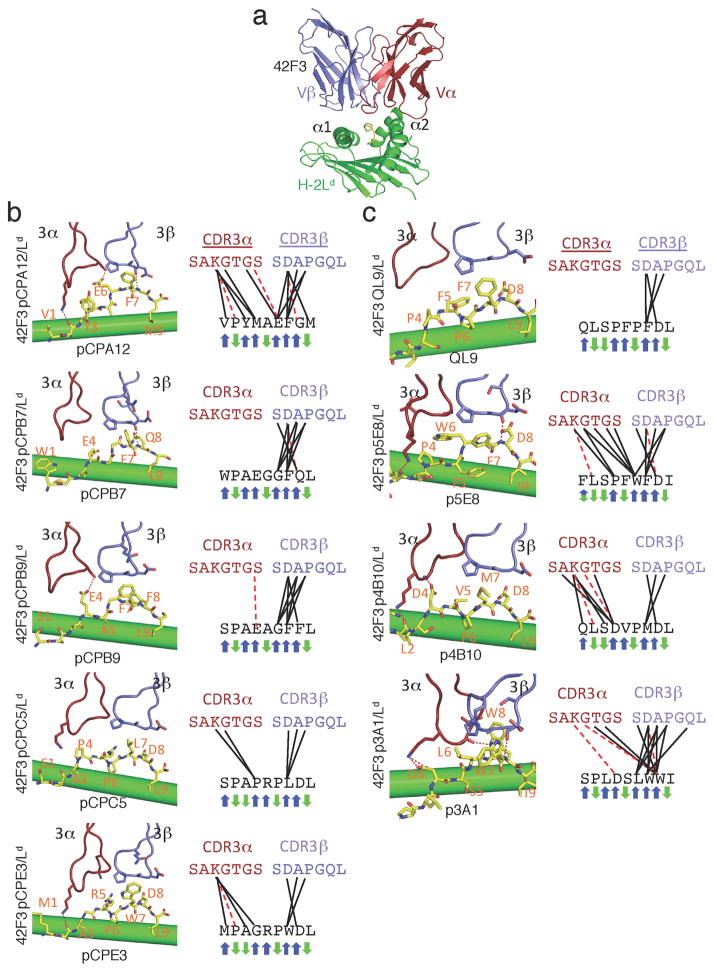
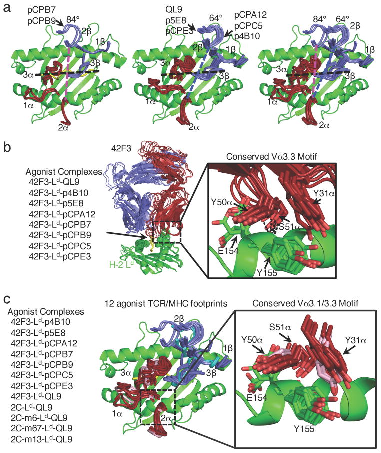
The T cell antigen receptor (TCR)-peptide-major histocompatibility complex (MHC) interface is composed of conserved and diverse regions, yet the relative contribution of each in shaping recognition by T cells remains unclear. Here we isolated cross-reactive peptides with limited homology, which allowed us to compare the structural properties of nine peptides for a single TCR-MHC pair. The TCR's cross-reactivity was rooted in highly similar recognition of an apical 'hot-spot' position in the peptide with tolerance of sequence variation at ancillary positions. Furthermore, we found a striking structural convergence onto a germline-mediated interaction between the TCR CDR1α region and the MHC α2 helix in twelve TCR-peptide-MHC complexes. Our studies suggest that TCR-MHC germline-mediated constraints, together with a focus on a small peptide hot spot, might place limits on peptide antigen cross-reactivity.


Conflict of interest statement

Competing financial interests

The authors declare no competing financial interests.

Figures

Fig. 1
Fig. 1
TCR selections of a circularly-permuted H-2Ld- yeast displayed library (a) Cartoon schematic of the circularly permuted mini-MHC m31r-CP and its presentation on the surface of EBY100 yeast as an Aga2 fusion protein. Inset is a stick schematic of the tethered QL9 peptide. Peptide positions are labeled accordingly and the general orientation of the TCR and MHC relative to the peptide is indicated with arrows. (b) The amino acid composition design of a ‘random’ peptide library where the P2 and P9 anchor positions are weighted to specific amino acids and the sequence of a natively displayed peptide QL9. (c) Representative peptides recovered by selection of first- and second-generation H-2Ld peptide libraries with 42F3 TCR multimers. Amino acids at a given position identical to those in QL9 are colored green. Amino acids with similar properties to QL9 are colored orange. Non-homologous resides are colored black. (d) The positional amino acid probabilities of peptides recovered from the enriched yeast populations specific for 42F3 TCR. Redundant peptide clones are represented once.
Fig. 2
Fig. 2
Signaling properties of synthetic peptide antigens (a) IL-2 response screen of the CD8+ (black bars) and CD8- (green bars) 42F3 T cells to APCs in the presence of 10μM synthesized peptide from first-generation (left) and second-generation (right) yeast displayed pMHC libraries. Data is represented as mean ± s.e.m. relative A450, normalized to anti-CD3 stimulation condition n=2 of technical replicates. (b) Scatterplots showing a wide range in affinities and activities, but little correlation between the IL-2 EC50 (left) or maximal IL-2 response (Emax) (right, as shown in Supplementary Fig. 2) of CD8+ 42F3 T with affinity of the 42F3-pMHC interactions. pCPE3 is a weak agonist for which EC50 and Emax were not determined, and was therefore omitted from this analysis.
Fig. 3
Fig. 3
Peptide specificity of the 42F3 TCR (a) The overall structure of the 42F3-QL9/H-2Ld complex. The MHC is illustrated in green, the TCRα in red, the TCRβ in blue and the peptide in yellow. (b-c) The peptide recognition by CDR3α and CDR3β loops of the 42F3 TCR for five synthetic peptides derived from the (b) second-generation yeast displayed library and (c) four from the first-generation library. Contacts are depicted as (left) structures and (right) contact maps. TCR residues making contacts to the peptides are represented as sticks. TCR CDR3 loops and the peptide identity are labeled in each panel. Black lines represent van der Waals contacts and red dashed lines represent hydrogen bonds. Blue (TCR-exposed) and green (MHC-buried) arrows illustrate the direction of the amino acid side chains on the TCR-pMHC complex.
Fig. 4
Fig. 4
Two agonist geometries accommodated by Vα3 germline motif (a) Overlaid geometric docking modes of the 42F3 TCR to H-2Ld binding at an 84° docking angle (left), 64° docking angle (center) and an overlay for comparison (right). The MHC is illustrated as a green ribbon, where the Vα CDR loops are in red and Vβ CDR loops are in blue. A black line is used to identify the QL9 peptide position and dashed colored lines are used to connect equivalent positions in CDR2α and CDR2β positions in the 84°(pink) and 64°(blue) geometries. Peptide names label the corresponding TCR-pMHC geometries. (b) The conserved germline contacts of the 42F3 Vα3.3 (red) to the H-2Ld α2 helix (green) across eight agonist complexes. The conserved germline contacts are inset and illustrated as sticks and dashed lines represent hydrogen bonds. (c) The conserved germline contacts of TCR Vα3 to the H-2Ld α2 helix (green) across twelve agonist complexes of 42F3 (red), 2C and 2C-variants (pink). The conserved germline contacts are illustrated as sticks and dashed lines represent hydrogen bonds.

References

    1. Adams JJ, Narayanan S, Liu B, Birnbaum ME, Kruse AC, Bowerman NA, et al. T Cell Receptor Signaling Is Limited by Docking Geometry to Peptide-Major Histocompatibility Complex. Immunity. 2011;35(5):681–693. - PMC - PubMed
    1. Rossjohn J, Gras S, Miles JJ, Turner SJ, Godfrey DI, McCluskey J. T cell antigen receptor recognition of antigen-presenting molecules. Annu Rev Immunol. 2015;33:169–200. - PubMed
    1. Beringer DX, Klejwegt FS, Wiede F, van der Slik AR, Loh KL, Petersen J, et al. T cell receptor reversed polarity recognition of a self-antigen major histocompatibility complex. Nat Immunol. 2015;16(11) - PubMed
    1. Dai S, Huseby ES, Rubtsova K, Scott-Browne J, Crawford F, Macdonald WA, et al. Crossreactive T Cells spotlight the germline rules for alphabeta T cell-receptor interactions with MHC molecules. Immunity. 2008;28(3):324–334. - PMC - PubMed
    1. Feng D, Bond CJ, Ely LK, Maynard J, Garcia KC. Structural evidence for a germline-encoded T cell receptor-major histocompatibility complex interaction ’codon’. Nat Immunol. 2007;8(9):975–983. - PubMed

Publication types

MeSH terms