

doi: 10.1107/S1600577515016604.
Epub 2015 Oct 3.
MASSIF-1: a beamline dedicated to the fully automatic characterization and data collection from crystals of biological macromolecules
Affiliations
- PMID: 26524320
- PMCID: PMC4629869
- DOI: 10.1107/S1600577515016604
Item in Clipboard
MASSIF-1: a beamline dedicated to the fully automatic characterization and data collection from crystals of biological macromolecules
J Synchrotron Radiat.
2015 Nov.
Abstract
MASSIF-1 (ID30A-1) is an ESRF undulator beamline operating at a fixed wavelength of 0.969 Å (12.8 keV) that is dedicated to the completely automatic characterization of and data collection from crystals of biological macromolecules. The first of the ESRF Upgrade MASSIF beamlines to be commissioned, it has been open since September 2014, providing a unique automated data collection service to academic and industrial users. Here, the beamline characteristics and details of the new service are outlined.
Keywords: automatic data collection; automation; beamline; radiation damage.
Figures
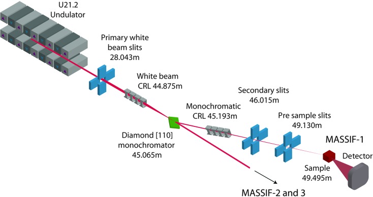
Schematic of the MASSIF-1 layout. Major components are shown with their distances from the source.
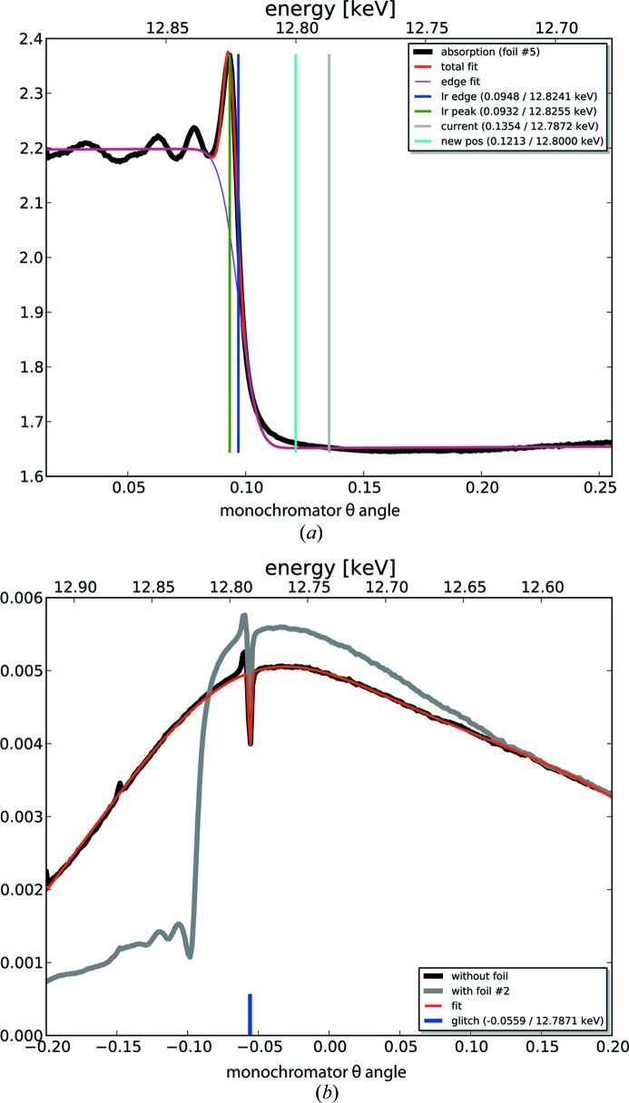
Calibration of the MASSIF-1 Laue [110] diamond monochromator. (a) Scan in transmission mode of the Ir L
II absorption edge. The blue line shows the position of the inflection point, the cyan line the angle to which the monochromator is moved to ensure a monochromatic X-ray beam of E = 12.80 keV (λ = 0.969 Å). (b) Scan of the MASSIF-2 monochromator θ angle; the glitch from the MASSIF-1 monochromator can be seen at 12.78 keV before it was moved to the calibrated position. Intensity units are arbitrary.
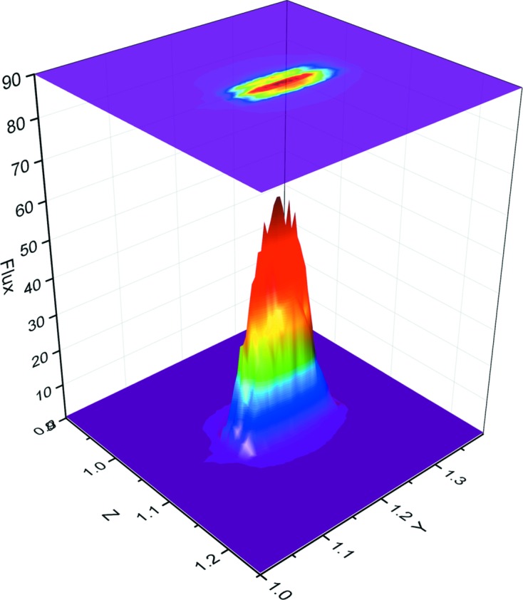
Three-dimensional profile of the beam at the sample position. The two-dimensional profile is shown above. The flux is shown in arbitrary units.
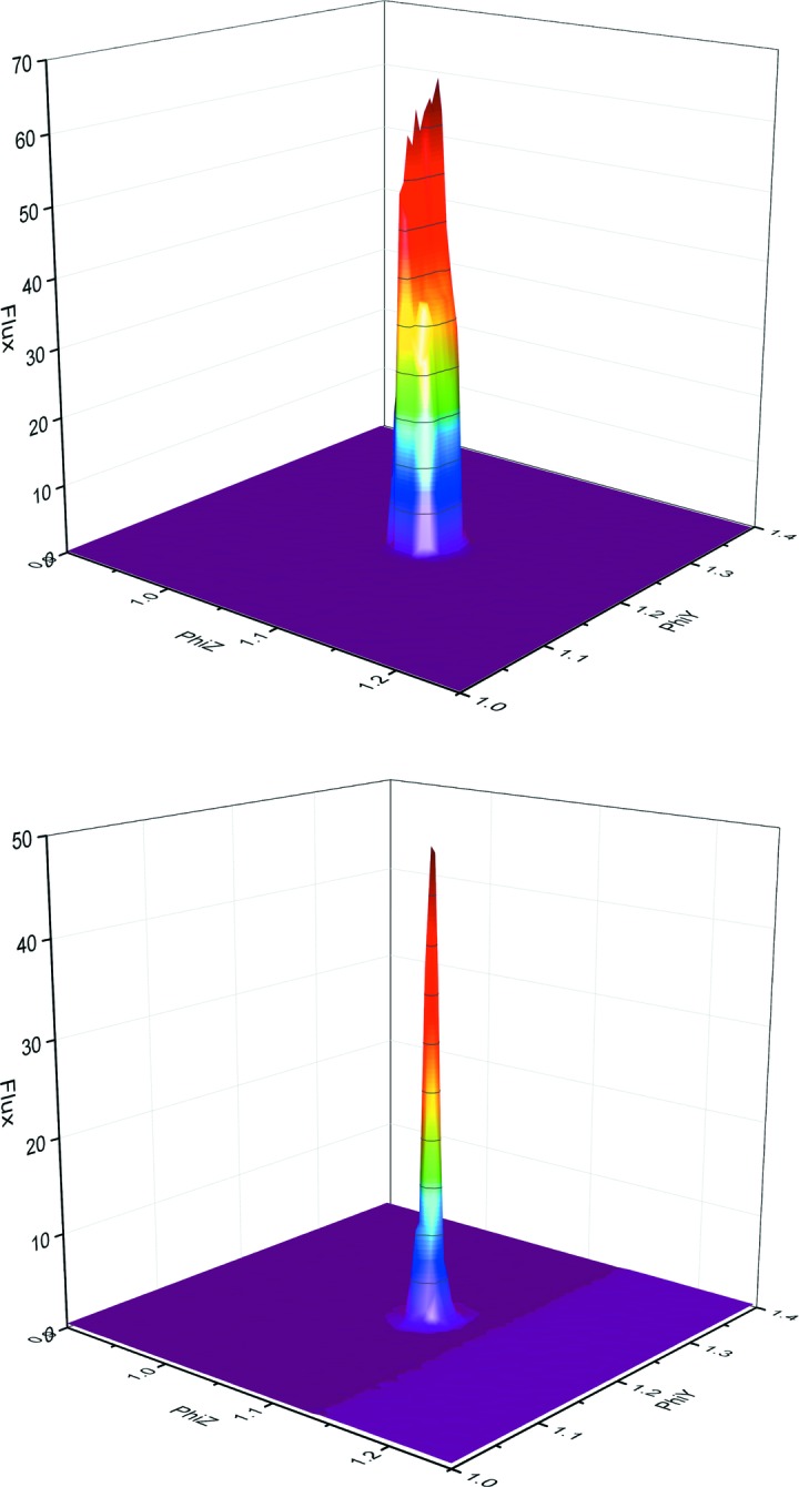
Three-dimensional profile of the beam when using apertures. The 50 µm (top) and 10 µm (below) profiles are shown; 30 µm, 20 µm and 15 µm apertures are also available. Using the central part of the full beam Gaussian leads to a near ‘top-hat’ profile of the beam. The flux is shown in arbitrary units.
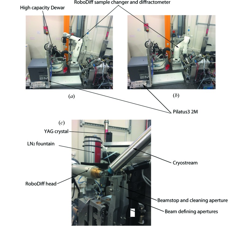
Experimental hutch configuration. The main components of the MASSIF-1 experimental hutch are shown with the RoboDiff in parked (a) and goniometer positions (b). (c) A close-up of the MASSIF-1 sample environment with the RoboDiff in goniometer position.
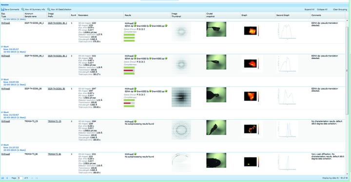
ISPyB summary page. A screenshot showing the display in ISPyB of results, such as diffraction maps, line scans for centring, centring snap shots, diffraction images and the results of autoprocessing, for a series of samples processed using the fully automatic protocols available at MASSIF-1. Comments are automatically written (far right column) to inform users on various stages of the process such as ‘weak diffraction’, default 180° data collection’ etc.
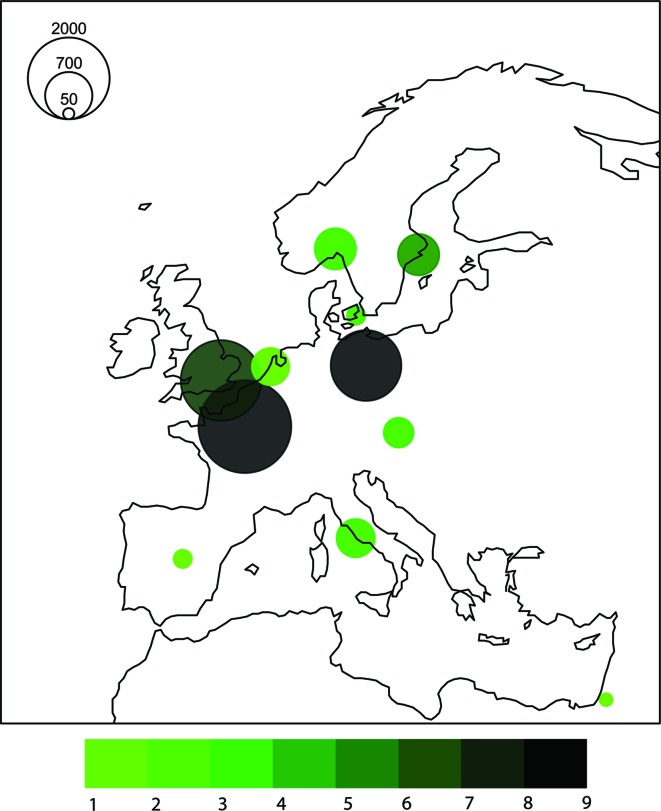
Distribution of crystals sent to MASSIF-1 from across Europe in the first eight months of operation (September 2014–June 2015). Circles are scaled to the number of crystals and are centred on the capital city of the country from which the crystals were sent. The colour is scaled to the number of groups using the beamline.
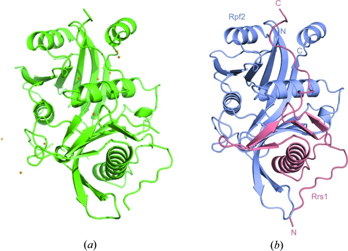
The Rpf2-Rrs1 complex structure solved using diffraction data collected automatically on MASSIF-1. (a) The model determined by the automatic SAD pipeline is shown as a cartoon from the Cα trace with the Se atom substructure shown as gold crosses. (b) The final refined model is shown with Rpf2 in magenta and Rrs1 in salmon.
References
-
- Bowler, M. W., Guijarro, M., Petitdemange, S., Baker, I., Svensson, O., Burghammer, M., Mueller-Dieckmann, C., Gordon, E. J., Flot, D., McSweeney, S. M. & Leonard, G. A. (2010). Acta Cryst. D66, 855–864. - PubMed
-
- Bowler, M. W., Mueller, U., Weiss, M., Sanchez-Weatherby, J., Sorensen, T., Thunnissen, M., Ursby, T., Gobbo, A., Russi, S., Bowler, M. G., Brockhauser, S., Svensson, O. & Cipriani, F. (2015). Cryst. Growth Des. 15, 1043–1054.
Publication types
MeSH terms
Substances
LinkOut - more resources
Full Text Sources
Other Literature Sources
