Review
. 2015 Nov 2;7(11):a019422.
doi: 10.1101/cshperspect.a019422.

Germline and Pluripotent Stem Cells

Affiliations
Review

Germline and Pluripotent Stem Cells

Wolf Reik et al. Cold Spring Harb Perspect Biol. .

Abstract

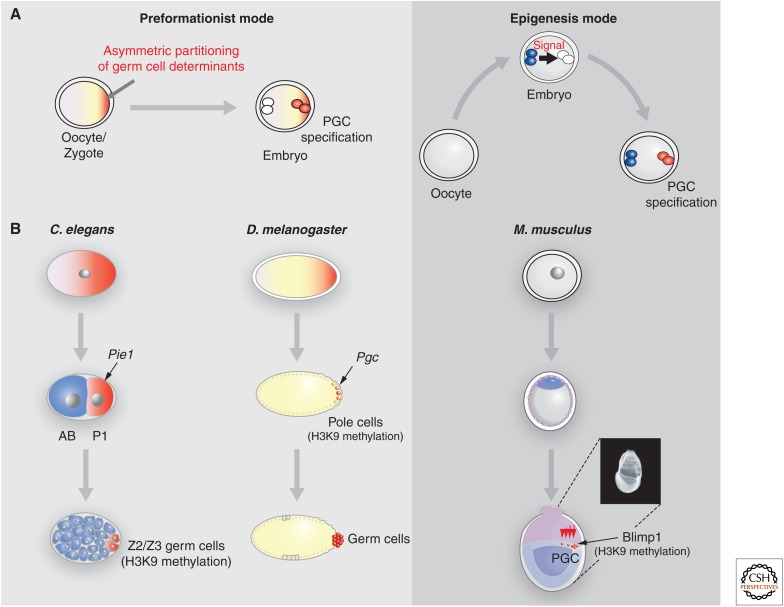
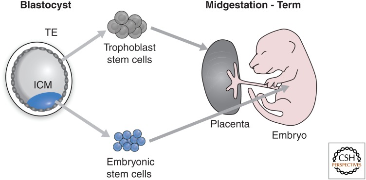
Epigenetic mechanisms play an essential role in the germline and imprinting cycle. Germ cells show extensive epigenetic programming in preparation for the generation of the totipotent state, which in turn leads to the establishment of pluripotent cells in blastocysts. The latter are the cells from which pluripotent embryonic stem cells are derived and maintained in culture. Following blastocyst implantation, postimplantation epiblast cells develop, which give rise to all somatic cells as well as primordial germ cells, the precursors of sperm and eggs. Pluripotent stem cells in culture can be induced to undergo differentiation into somatic cells and germ cells in culture. Understanding the natural cycles of epigenetic reprogramming that occur in the germline will allow the generation of better and more versatile stem cells for both therapeutic and research purposes.


Figures

Figure 1.
Figure 1.
Potential of the mammalian oocyte, zygote, and blastocyst. (A) The mammalian oocyte contains maternal RNAs and proteins (maternal inheritance), which can determine early developmental events, genetic information (maternal chromosomes), and epigenetic information (DNA methylation and chromatin marks). (B) The zygote gives rise to the blastocyst with its inner cell mass (ICM) cells (blue) giving rise to ES cells in culture. The epiblast derivative of the ICM in the postimplantation blastocyst gives rise to all somatic cells and PGCs. A range of pluripotent stem cells (top line) can be derived from the various cell types isolated from early- and late-stage blastocysts and later primitive streak embryos. Types of stem cell include XEN, extraembryonic endoderm; ES, embryonic stem; TS, trophoblast stem; EpiSC, epiblast stem cell; EG, embryonic germ.
Figure 2.
Figure 2.
The epigenetic reprogramming cycle in mammalian development. Immediately after fertilization in the zygote, the paternal pronucleus (PN) is packaged with histones that lack H3K9me2 and H3K27me3, whereas the maternal chromatin contains these marks. The paternal PN also rapidly loses 5-methylcytosine (5mC) on a genome-wide scale, whereas the maternal does not. Passive loss of 5mC occurs during preimplantation development until the blastocyst stage when the ICM cells begin to acquire high levels of 5mC, H3K9me2, and H3K27me3. The placenta, which is largely derived from the TE of the blastocyst, remains relatively hypomethylated. PGCs undergo demethylation of 5mC and H3K9me2 progressively as they migrate into the gonads. De novo DNA methylation, including parent-specific imprinting, takes place during gametogenesis.
Figure 3.
Figure 3.
Early germ cell determination in the mouse. (A) Two models summarize the mode by which germ cells are determined in various organisms. The preformationist mode assumes one or more localized determinants in the oocyte or early embryo specify progeny cells becoming PGCs. In the epigenesis mode, a signal emanating from a neighboring cell(s) in the early embryo determines the future PGCs. (B) This part of the figure highlights the features that contribute to the repression of somatic gene programs during germline specification in various organisms. In Caenorhabditis elegans, the germline lineage (red) is specified after the first division of the zygote by expression of Pie1, which confers transcriptional quiescence. The other cell (blue) gives rise to somatic tissues. In Drosophila melanogaster, the precursors of the germ cells are the so-called pole cells contained on one side of the zygote syncytium (i.e., multinucleated); transcriptional quiescence in these cells depends on localized RNA from the gene Pgc and high levels of H3K9 methylation. In Mus musculus, the earliest precursors of the germ cells are visible by expression of Blimp1 at the base of the allantois. Blimp1 initiates transcriptional quiescence in these cells.
Figure 4.
Figure 4.
Germline development in mice. Postimplantation proximal competent epiblast cells at E6 (pink) respond to BMP4 signal from the extraembryonic tissues (pink), which activates BLIMP1. Expression of BLIMP1 marks the onset of commitment to the primordial germ cell fate (red), whereas other cells become somatic cells.
Figure 5.
Figure 5.
Early epigenetic events during germ cell specification. Expression of Blimp1, Prdm14, and Tcfap2c in descendants of epiblast cells leads to repression of the somatic gene expression program and initiation of the germ cell program (red). This is followed by expression of Stella, Nanog, and Esg1, increase in the H3K4me3 and H3K9ac active marks, as well as the repressive H3K27me3 mark (*), and loss of H3K9me2 and 5mC. The PGCs start to show loss of DNA methylation as they migrate to the developing gonads, with comprehensive loss of DNA methylation and the erasure of imprints occurring shortly after they enter the gonads. PRDM9 is crucial in the later process of gametogenesis, marking the transition from PGCs to gametes. This occurs with the onset of meiosis at E13.5 in females. **In males gametogenesis occurs postnatally.
Figure 6.
Figure 6.
Differentiation of ES cells into different cell types in vitro. ES cells can be differentiated in vitro under suitable culture conditions into many different cell types such as neurons, muscle cells, and even germ cells (oocytes).
Figure 7.
Figure 7.
ES and TS cells from the blastocyst. ES cells are derived from ICM cells and can be kept in culture without differentiating. They can be genetically manipulated while in culture. ES cells can be reintroduced into blastocysts and then colonize all tissues in the embryo, including the germline, but excluding the TS cells of the placenta. TS cells can be established similarly into culture from the TE cells of the blastocyst and, when reintroduced into blastocysts, contribute to placental cell types.
Figure 8.
Figure 8.
Pluripotent cells have the capacity to reprogram somatic cells. ES or EG cells can be fused with somatic cells, resulting in tetraploid hybrids. This leads to epigenetic reprogramming of the somatic nucleus, with changes in, for example, 5MeC, H3 and H4 acetylation, and H3K4 methylation. The tetraploid cells resulting from this fusion or reprogramming that occurs when producing iPSCs also have a pluripotent phenotype: When injected into blastocysts, they can contribute to many different cell types in the embryo.

References

    1. Allis CD, Jenuwein T, Reinberg D. 2014. Overview and concepts. Cold Spring Harb Perspect Biol 10.1101/cshperspect.a018739. - DOI
    1. Ancelin K, Lange UC, Hajkova P, Schneider R, Bannister AJ, Kouzarides T, Surani MA. 2006. Blimp1 associates with Prmt5 and directs histone arginine methylation in mouse germ cells. Nat Cell Biol 8: 623–630. - PubMed
    1. Arney KL, Bao S, Bannister AJ, Kouzarides T, Surani MA. 2002. Histone methylation defines epigenetic asymmetry in the mouse zygote. Int J Dev Biol 46: 317–320. - PubMed
    1. Avilion AA, Nicolis SK, Pevny LH, Perez L, Vivian N, Lovell-Badge R. 2003. Multipotent cell lineages in early mouse development depend on SOX2 function. Genes Dev 17: 126–140. - PMC - PubMed
    1. Barlow DP, Bartolomei MS. 2014. Genomic imprinting in mammals. Cold Spring Harb Perspect Biol 6: a018382. - PMC - PubMed

LinkOut - more resources