Review
. 2015 Nov 2;7(11):a021733.
doi: 10.1101/cshperspect.a021733.

The Corticohippocampal Circuit, Synaptic Plasticity, and Memory

Affiliations
Review

The Corticohippocampal Circuit, Synaptic Plasticity, and Memory

Jayeeta Basu et al. Cold Spring Harb Perspect Biol. .

Abstract

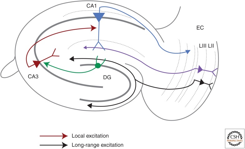
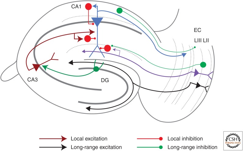
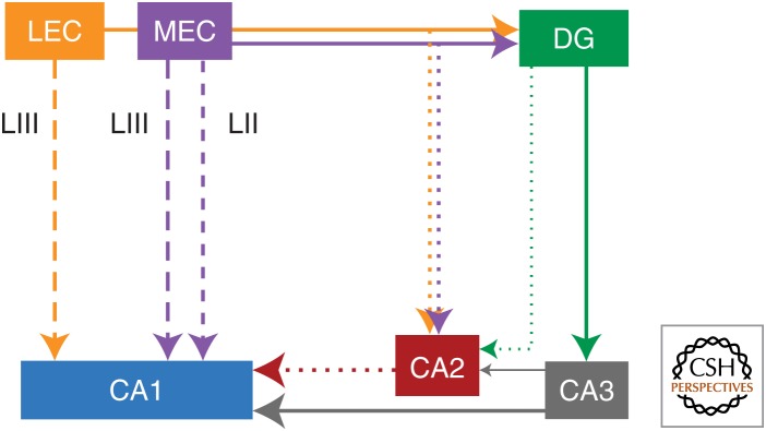
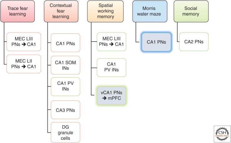
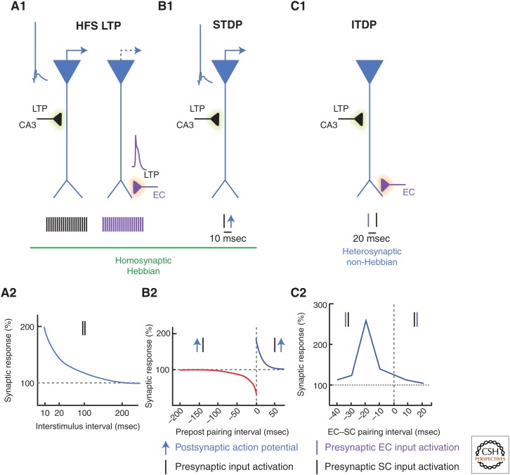
Synaptic plasticity serves as a cellular substrate for information storage in the central nervous system. The entorhinal cortex (EC) and hippocampus are interconnected brain areas supporting basic cognitive functions important for the formation and retrieval of declarative memories. Here, we discuss how information flow in the EC-hippocampal loop is organized through circuit design. We highlight recently identified corticohippocampal and intrahippocampal connections and how these long-range and local microcircuits contribute to learning. This review also describes various forms of activity-dependent mechanisms that change the strength of corticohippocampal synaptic transmission. A key point to emerge from these studies is that patterned activity and interaction of coincident inputs gives rise to associational plasticity and long-term regulation of information flow. Finally, we offer insights about how learning-related synaptic plasticity within the corticohippocampal circuit during sensory experiences may enable adaptive behaviors for encoding spatial, episodic, social, and contextual memories.


Figures

Figure 1.
Figure 1.
The classical corticohippocampal glutamatergic circuits. The classical corticohippocampal circuit comprises of glutamatergic input from the superficial entorhinal cortex (EC) layers (LII and LIII) to CA1 pyramidal neurons via the trisynaptic and monosynaptic paths; hippocampal back projections to the deep layers of EC complete the loop. Sensory signals drive the perforant path (PP, purple) inputs from EC LIII pyramidal neurons to distal CA1 pyramidal neuron dendrites (light blue). Activated EC LII pyramidal neurons send inputs to dentate gyrus (DG, black), which sends mossy fiber axons (dark green) to CA3 and then CA3 feeds onto CA1 neurons through Schaffer collateral (SC, dark red) excitatory inputs. A major output of the hippocampus arises from CA1 pyramidal neurons, which project to lateral ventricles (LVs) of EC. There is a 15-20 ms timing delay for transmission of information from EC LII to CA1 through the trisynaptic path compared with that from EC LIII to CA1 via the monosynaptic path.
Figure 2.
Figure 2.
Local and long-range GABAergic connections. CA1 has several local GABAergic interneurons (red). These target the CA1 pyramidal neuron soma, axon, and dendrite to modulate pyramidal neuron activity in a domain-specific manner. Schaffer collateral (SC)- and entorhinal cortex (EC)-associated inhibitory microcircuits provide feedforward inhibition, whereas feedback inhibition is recruited recurrently when the CA1 pyramidal neuron fires an action potential. Long-range inhibitory projections (green) from the EC provide direct inhibition preferentially to local interneurons (INs) in CA1. Long-range projections from GABAergic neurons in stratum oriens (SO) of hippocampus to layer II/III (LII/LIII) of the EC have also been described.
Figure 3.
Figure 3.
The updated corticohippocampal circuit. This circuit diagram integrates the recently discovered glutamatergic inputs from the entorhinal cortex (EC) to the hippocampus as well as connections within the hippocampus. In addition to the classical trisynaptic (EC layer II [LII] → dentate gyrus [DG] → CA3 → CA1, solid line) and monosynaptic (EC layer III (LIII) → CA1, large-dashed lines) pathways of information flow, CA1 also receives monosynaptic projections from LII of the medial entorhinal cortex (MEC) (small dashed line) and CA2 receives direct inputs from LII of both MEC and lateral entorhinal cortex (LEC) (dotted lines). Within the hippocampus, CA2 sends prominent inputs to CA1, targeting dendritic domains (stratum oriens [SO]/stratum radiatum [SR]) (red) that overlap with the CA3 → CA1 inputs. CA2 also receives weak inputs from DG and CA3. The thickness of the arrowed lines emphasizes the strength of the input connection.
Figure 4.
Figure 4.
Cellular and circuit correlates of behavioral learning. Genetic and classical lesions have elucidated how the different pathways and cell populations comprising the corticohippocampal circuit support various forms of associational learning and declarative memory functions. All subfields, except the grayed boxes, show results from behavioral experiments involving genetically targeted cell type or input specific functional manipulations. Gray boxes indicate physical or chemical lesion-based findings. Both the medial entorhinal cortex (MEC) layer II (LII) (Kitamura et al. 2014) and layer III (LIII) (Suh et al. 2011) glutamatergic inputs to CA1 are used for trace fear conditioning (TFC). CA1 pyramidal neurons (PNs) (Goshen et al. 2011), CA3 pyramidal neurons (Nakashiba et al. 2008), dentate gyrus (DG), granule cells (Nakashiba et al. 2012; Kheirbek et al. 2013), parvalbumin (PV) interneurons (INs) (Donato et al. 2013), and somatostatin (SOM) interneurons (Lovett-Barron et al. 2014) have all been found to be involved in contextual fear-learning behavior. Spatial working memory requires activity in the MEC LIII pyramidal neurons projections to CA1 (Suh et al. 2011; Yamamoto et al. 2014), CA1 PV interneurons (Murray et al. 2011), and the ventral hippocampal projections to the prefrontal cortex (Wang and Cai 2006).
Figure 5.
Figure 5.
Synaptic learning motifs and their temporal fidelity. Scheme showing how different synaptic learning rules emerge in the CA1 microcircuit from temporally patterned activity of synaptic inputs (CA3 to CA1 Schaffer collateral [SC] inputs in black; entorhinal cortex [EC] to CA1 inputs in purple) and the postsynaptic CA1 pyramidal neuron (blue). (A1) High-frequency stimulation long-term potentiation (HFS LTP) is a homosynaptic form of synaptic learning in which strong tetanic stimulation (200 pulses at 20–100 Hz) of the CA3 or EC inputs can strengthen the synaptic output of that specific active input pathway. HFS LTP is Hebbian in that its induction requires Schaffer collateral (SC) input stimulation to evoke somatic spikes, or perforant path (PP) input stimulation to trigger dendritic spikes in the postsynaptic CA1 pyramidal neurons. (A2) Plot depicting the frequency dependence of SC HFS LTP. Postsynaptic excitatory response recorded in CA1 pyramidal neurons plotted as a function of the interstimulus interval for tetanic stimulation of the CA3–CA1 SC inputs. Preinduction baseline synaptic response is 100%. (Adapted, with values, from Thomas et al. 1996; Aihara et al. 1997; Zakharenko et al. 2003; Alarcon et al. 2004.) (B1) Spike-timing-dependent plasticity (STDP) is induced in CA1 pyramidal neurons by temporally precise pairing of synaptic inputs from CA3 with a postsynaptic spike triggered by injecting a brief current step in the soma. The pairing is typically repeated 50–100 times at 10 Hz. (B2) LTP is induced when the presynaptic input precedes the postsynaptic spike by 5–20 msec, whereas long-term depression (LTD) prevails when the pairing sequence is reversed (postsynaptic spike before the presynaptic input). (−) Timing intervals indicate pre- before postsynaptic pairing. (Adapted from data in Bi and Poo 1998; Debanne et al. 1998; Nishiyama et al. 2000.) (C1) Input-timing-dependent plasticity (ITDP) is induced when EC and SC inputs are stimulated 20 msec apart (EC before SC) at subthreshold strengths (hence, non-Hebbian) for 90 sec at a 1 Hz frequency. ITDP is expressed in the CA1 pyramidal neuron as a long-term potentiation (LTP) of the SC- mediated postsynaptic depolarization without a change in the PP-evoked response (hence, heterosynaptic). (C2) Induction of ITDP is finely tuned to the 20 msec pairing interval, even a 10 msec deviation from this preferred timing interval is ineffective. (−) Timing intervals indicate EC before SC input pairing. (Adapted from data in Basu et al. 2013.)

References

    1. Abel T, Nguyen PV, Barad M, Deuel TA, Kandel ER, Bourtchouladze R. 1997. Genetic demonstration of a role for PKA in the late phase of LTP and in hippocampus-based long-term memory. Cell 88: 615–626. - PubMed
    1. Abraham WC, Tate WP. 1997. Metaplasticity: A new vista across the field of synaptic plasticity. Prog Neurobiol 52: 303–323. - PubMed
    1. Ahmed OJ, Mehta MR. 2009. The hippocampal rate code: Anatomy, physiology and theory. Trends Neurosci 32: 329–338. - PMC - PubMed
    1. Aihara T, Tsukada M, Crair MC, Shinomoto S. 1997. Stimulus-dependent induction of long-term potentiation in CA1 area of the hippocampus: Experiment and model. Hippocampus 7: 416–426. - PubMed
    1. Alarcon JM, Hodgman R, Theis M, Huang YS, Kandel ER, Richter JD. 2004. Selective modulation of some forms of Schaffer collateral-CA1 synaptic plasticity in mice with a disruption of the CPEB-1 gene. Learn Mem 11: 318–327. - PMC - PubMed

LinkOut - more resources