

Review
doi: 10.1152/physiol.00030.2015.
ASICs Mediate Pain and Inflammation in Musculoskeletal Diseases
Affiliations
- PMID: 26525344
- PMCID: PMC4747907
- DOI: 10.1152/physiol.00030.2015
Item in Clipboard
Review
ASICs Mediate Pain and Inflammation in Musculoskeletal Diseases
Physiology (Bethesda).
2015 Nov.
Abstract
Chronic musculoskeletal pain is debilitating and affects ∼ 20% of adults. Tissue acidosis is present in painful musculoskeletal diseases like rheumatoid arthritis. ASICs are located on skeletal muscle and joint nociceptors as well as on nonneuronal cells in the muscles and joints, where they mediate nociception. This review discusses the properties of different types of ASICs, factors affecting their pH sensitivity, and their role in musculoskeletal hyperalgesia and inflammation.
©2015 Int. Union Physiol. Sci./Am. Physiol. Soc.
Conflict of interest statement
No conflicts of interest, financial or otherwise, are declared by the author(s).
Figures
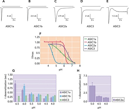
Acid-evoked currents and biophysical properties of homomeric ASICs A–E: CHO-K1 cells transfected with homomeric ASICs were bathed in ringer solution and activated by rapid application of a pH 4 solution. F: proton affinities (pH sensitivity) for homomeric ASICs were measured by lowering the extracellular pH from 7.4 to 4. G and H: the rate of desensitization (τ desensitization) of ASICs was measured at different extracellular pH values and demonstrated that desensitization of all ASIC subunits except ASIC3 is increased when pH is lowered. Figure was adapted from Ref. , with permission from The Journal of Biological Chemistry.
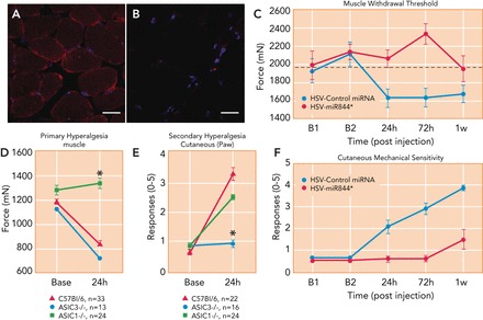
ASICs are expressed on muscle tissue and play a role in carrageenan-induced inflammatory muscle pain A and B: representative muscle tissue sections from wild-type (A) and ASIC3−/− (B) mice, showing ASIC3 (red) and DAPI (nuclear stain, blue) staining. Figure reproduced from Ref. with permission. C: genetic knockdown of ASIC3 in primary afferent fibers innervating muscle, using HSV-miRNA to ASIC3 (red, HSV-miR844), prevent the development of primary muscle hyperalgesia induced by intramuscular carrageenan injection compared with animals treated with control HSV-miRNA (blue). *Significantly greater than controls (P < 0.05). Reproduced from Ref. with permission. D: muscle withdrawal thresholds (primary hyperalgesia) before and after induction of muscle inflammation in WT (red), ASIC3−/− (blue), and ASIC1−/− (green) mice. ASIC1−/− do not develop muscle hyperalgesia 24 h after inflammation with carrageenan compared with WT and ASIC3−/− mice. *Significantly greater than WT and ASIC3−/− (P < 0.05). Reproduced from Ref. with permission. E: responses to repeated mechanical stimulation of the paw (secondary hyperalgesia) before and after induction of muscle inflammation in WT (red), ASIC3−/− (blue), and ASIC1−/− (green) mice. ASIC3−/− do not develop secondary hyperalgesia 24 h after inflammation with carrageenan compared with WT and ASIC1−/− mice. Reproduced from Ref. with permission. F: genetic knockdown of ASIC3 in primary afferent fibers innervating muscle, using HSV-miRNA to ASIC3 (red, HSV-miR844), prevents development of secondary hyperalgesia induced by intramuscular carrageenan compared with animals treated with control HSV-miRNA (blue). *Significantly greater than controls (P < 0.05). Reproduced from Ref. with permission.
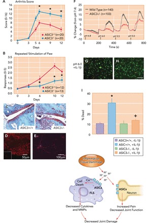
ASIC3 mediates the nociception produced by collagen-antibody-induced arthritis, while protecting the joint from inflammation and destruction A: ASIC3−/− mice (blue) show enhanced inflammation after induction of collagen antibody-induced arthritis (CAIA) compared with WT mice (red). *Significantly greater than controls (P < 0.05). Reproduced from Ref. with permission. B: CAIA-induced secondary hyperalgesia of the paw (number of responses to repeated mechanical stimuli) was attenuated in the ASIC3−/− mice (blue) compared with wild-type mice (red). *Significantly lower than wild-type mice (P < 0.05). Reproduced from Ref. with permission. C: representative sections from the ankle joint of animals 12 days after induction of CAIA stained with Safronin-0 from wild-type (ASIC3+/+) and ASIC3−/− mice. Notice enhanced inflammation and damage in tissue section from an ASIC3−/− mouse. Reproduced from Ref. with permission. D: fluorescent micrograph from the synovium of the knee joint immunohistochemically stained for ASIC3 in wild-type mice. Inset shows ASIC3 immunohistochemistry in synovium from ASIC3−/− mice. Reprinted from Ref. with permission. E: fluorescent immunohistochemical staining for ASIC3 (red) in cultured fibroblast-like synovocytes (FLS). Nuclei are stained with TO-PRO3 (blue). Reprinted from Ref. with permission. F: intracellular calcium concentration ([Ca]2+) was calculated in wild-type (black) and ASIC3−/− (red) FLS, loaded with [Ca]2+-sensitive fluorescent indicator Oregon Green BAPTA-1 AM (OGB-1), in response to exposure to decreasing pH values from 6.8 to 5 (150-s incubation). The response was normalized as percentage change from pH 7.4. Reproduced from Ref. with permission. G and H: live/dead assay of cells of cultured FLS extracted from wild-type (G) and ASIC3−/− (H) mice. FLS were treated with pH 6 after prior incubation with IL-1β. Green staining represents live cells, whereas red staining represents dead cells. Reproduced and modified with permission from Ref. . I: the average % dead cells after treating FLS cells with pH 6 and IL-1β in wild-type (red) and ASIC3−/− (blue) FLS. Notice a significant increase in the number of dead cells in wild-type mice treated with pH 6 and IL-1b (*) that did not occur in ASIC3−/− mice treated with pH 6 and IL-1b (+). Reproduced and modified with permission from Ref. . J: schematic presentation for the protective role of ASIC3 in collagen-induced arthritis model (CAIA). In this model, the acidic enviroment assocaited with inflammation activates ASIC3 on FLS. Activation of ASIC3 enhances intracellular Ca2+ release, leading to cell death, which limits the secretion of inflammatory cytokines and metalloproteases (MMPs) by FLS and limits joint damage (42). ASIC3 located on neurons is also activated by decreases in pH associated with inflammation, which increases pain limiting joint function, to ultimately decrease joint damage. The red arrows show the proposed pathways for activation of ASICs in FLS and in neurons, and how they ultimately lead to reduced joint damage.
References
-
- Akopian AN, Chen CC, Ding Y, Cesare P, Wood JN. A new member of the acid-sensing ion channel family. Neuroreport 11: 2217–2222, 2000. - PubMed
-
- Andersson G, and American Academy of Orthopaedic Surgeons. The Burden of Musculoskeletal Diseases in the United States: Prevalence, Societal, and Economic Cost. Rosemont, IL: American Acad. of Orthopaedic Surgeons, 2008.
-
- Askwith CC, Wemmie JA, Price MP, Rokhlina T, Welsh MJ. Acid-sensing ion channel 2 (ASIC2) modulates ASIC1 H+-activated currents in hippocampal neurons. J Biol Chem 279: 18296–18305, 2004. - PubMed
Publication types
MeSH terms
Substances
Grants and funding
LinkOut - more resources
Full Text Sources
Other Literature Sources
Medical