Review
. 2015 Nov 2;5(11):a021568.
doi: 10.1101/cshperspect.a021568.

Hepatitis D Virus Replication

Affiliations
Review

Hepatitis D Virus Replication

John M Taylor. Cold Spring Harb Perspect Med. .

Abstract

This work reviews specific related aspects of hepatitis delta virus (HDV) reproduction, including virion structure, the RNA genome, the mode of genome replication, the delta antigens, and the assembly of HDV using the envelope proteins of its helper virus, hepatitis B virus (HBV). These topics are considered with perspectives ranging from a history of discovery through to still-unsolved problems. HDV evolution, virus entry, and associated pathogenic potential and treatment of infections are considered in other articles in this collection.


Figures

Figure 1.
Figure 1.
Three HDV RNAs that accumulate during replication. The numbering of nucleotides refers to the 1679 genomes sequenced by Kuo et al. (1988b). The circular RNA genome and its exact complement, the antigenome, are drawn as unbranched rod-like structures. Both of these RNAs contain a small domain, indicated by a solid green box, which, after appropriate folding, will act as an efficient self-cleaving ribozyme. The antigenome contains a region, indicated by a solid brown box, that contains the open reading frames (ORFs) for the small and large delta antigens. However, these proteins are translated from a shorter linear RNA. This third RNA contains a 5′-cap structure and in part, because of an AAUAAA polyadenylation signal indicated by a small open green box, is 3′-polyadenylated. The ORF of the small delta antigen is increased to that of the large delta antigen, by a posttranscriptional RNA editing event that occurs at position 1014 of the antigenome, corresponding to the middle of an UAG amber termination codon, thereby allowing an extended translation via a UGG codon for tryptophan. In the liver of an infected animal, the abundances of these three RNAs per average cell are ∼300,000, 50,000, and 600 (Chen et al. 1986).
Figure 2.
Figure 2.
Two forms of the delta antigen. As a consequence of site-specific RNA editing during HDV RNA replication, the coding region for the small delta antigen (195 amino acids [aa]) is increased to allow translation of the large delta antigen (214 aa). The small and large forms are both highly basic proteins and share features, such as a nuclear localization signal and a region with a propensity to form α-helices. The unique carboxyl terminus of the large protein contains a cysteine, four amino acids from the terminus, which is the site for a farnesylation that, in turn, is essential for the ability of this protein to facilitate assembly using the envelope proteins of HBV.
Figure 3.
Figure 3.
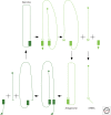
A rolling-circle model for the replication of the HDV RNA genome. This is one form of a double-rolling circle model, which uses both data and some speculations to explain how HDV RNA–directed RNA synthesis, using redirection of one or more host RNA polymerases, can produce multimeric transcripts, which, because of their ribozymes (filled rectangles), then undergo self-cleavage and ligation to produce new unit-length circular RNAs (Taylor et al. 2013). This model also tries to explain how some RNA transcripts of antigenomic polarity can go on to be processed to produce the observed mRNA for the delta antigens. Such a transcript is consistent with data that the 5′-end has a unique location and is capped, and that the 3′-end is polyadenylated as directed by an essential AAUAAA signal (open rectangle).

References

    1. Alves C, Freitas N, Cunha C. 2008. Characterization of the nuclear localization signal of the hepatitis delta virus antigen. Virology 370: 12–21. - PubMed
    1. Alves C, Cheng H, Roder H, Taylor J. 2010. Intrinsic disorder and oligomerization of the hepatitis delta virus antigen. Virology 407: 333–340. - PMC - PubMed
    1. Bergmann KF, Cote PJ, Gerin JL. 1990. Immunological characterization of hepatitis delta antigen. In Progress in clinical and biological research (ed. Gerin JL, Purcell RH, Rizetto M), pp. 165–171. Wiley, New York. - PubMed
    1. Bichko VV, Taylor JM. 1996. Redistribution of the delta antigens in cells replicating the genome of hepatitis delta virus. J Virol 70: 8064–8070. - PMC - PubMed
    1. Bichko V, Barik S, Taylor J. 1997. Phosphorylation of the hepatitis delta virus antigens. J Virol 71: 512–518. - PMC - PubMed

MeSH terms

LinkOut - more resources