. 2015 Nov 18;88(4):704-19.
doi: 10.1016/j.neuron.2015.10.005. Epub 2015 Oct 29.

Doublecortin-Like Kinases Promote Neuronal Survival and Induce Growth Cone Reformation via Distinct Mechanisms

Affiliations

Doublecortin-Like Kinases Promote Neuronal Survival and Induce Growth Cone Reformation via Distinct Mechanisms

Homaira Nawabi et al. Neuron. .

Abstract

After axotomy, neuronal survival and growth cone re-formation are required for axon regeneration. We discovered that doublecortin-like kinases (DCLKs), members of the doublecortin (DCX) family expressed in adult retinal ganglion cells (RGCs), play critical roles in both processes, through distinct mechanisms. Overexpression of DCLK2 accelerated growth cone re-formation in vitro and enhanced the initiation and elongation of axon re-growth after optic nerve injury. These effects depended on both the microtubule (MT)-binding domain and the serine-proline-rich (S/P-rich) region of DCXs in-cis in the same molecules. While the MT-binding domain is known to stabilize MT structures, we show that the S/P-rich region prevents F-actin destabilization in injured axon stumps. Additionally, while DCXs synergize with mTOR to stimulate axon regeneration, alone they can promote neuronal survival possibly by regulating the retrograde propagation of injury signals. Multifunctional DCXs thus represent potential targets for promoting both survival and regeneration of injured neurons.


Figures

Figure 1:
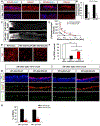
Figure 1:. DCLK2 expression is down-regulated by axotomy and DCLK2 overexpression promotes neuronal survival.
(A) Representative confocal images showing mRNA signal of DCLK1 and DCLK2 (red) in intact retina or those at 3 days post crush (dpc). Nuclei are labeled with DAPI (blue). Scale bar: 20μm. (B) Quantification of fluorescence intensity of DCLK1 and DCLK2 signals from (A). ***: p<0.001 (n=6). (C) Representative confocal images of optic nerve sections from wild type (WT) mice injected with AAV2-PLAP or AAV2-DCLK2. Axons are labeled with CTB. Scale bar: 100μm. (D) Quantification of the number of regenerating axons presented in (C). ***: p<0.001 (n=5). (E) Representative images of Tuj1-immunostained whole mount retinas 2 weeks post injury from wild type mice injected with AAV2-PLAP or AAV2-DCLK2. Scale bar: 20μm (F) Quantification of RGC survival as measured by Tuj1 staining. ***: p<0.001. (G) Representative confocal images of retinal sections taken at 24h or 72h post injury stained with antibodies against phospho-c-Jun (green), Tuj1 (blue) and probed by FISH with antisense probes of DCLK2 (red). Scale bar: 20μm. (H) Quantification of percentage of phospho-c-Jun-positive RGCs from (G). ***: p<0.001.
Figure 2:
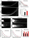
Figure 2:. DCLK2 enhances the initiation and elongation of optic nerve regeneration in PTEN-deleted mice.
(A) Representative confocal images of optic nerve sections taken 2 weeks after injury from PTENf/f mice injected with AAV2-Cre as well as AAV2-PLAP or AAV2-DCLK2. The red stars indicate the crush site. (B) Quantification of regenerative axons in the two groups. ***p<0.001 (n=7). (C) Quantification of RGC survival at 2 weeks after injury in PTENf/f mice injected with AAV2-Cre as well as AAV2-PLAP or AAV2-DCLK2. ***: p<0.001 (n=3). (D, E) Representative confocal images of optic nerve sections taken from 3 days (D) or 7 days (E) after injury from PTENf/f mice injected with AAV2-Cre as well as AAV2-PLAP or AAV2-DCLK2. (F, G) Quantification of the number of regenerative axons 3 days (F) and 1 week after injury (G). *p<0.05, ***p<0.001. At least 3 animals for each group. (H) Estimated axon regrowth rates (mm/week) in the two groups. ***: p<0.001, *: p<0.05. Scale bars: 100μm.
Figure 3:
Figure 3:. DCLK2 promotes growth cone formation.
(A) Time line of the experimental procedure. (B) Micrographs of neurites from retinal explants from the PTENf/f mice injected with AAV2-Cre and AAV2-PLAP or AAV2-DCLK2 stained with anti-DCLK2 (green), anti-Tuj1 (blue) and phalloidin (red). Scale bar: 5μm. (C) Quantification of DCLK2 fluorescence intensity normalized by volume of the neurite determined by phalloidin staining. ***: p<0.001. (D) Time-lapse micrographs of post-cut behavior of neurites in two groups for 1h after laser axotomy. Neurites are pseudo-colored in orange and the cut site is indicated by green arrowhead. Scale bar: 5μm. (E) Percentage of growth cone-forming neurites within 60 min after cut in explants from the two groups. ***: p<0.001 (n= 16-18 per group). (F) Quantification of distances navigated by the growth cones from the retraction site in the two groups during the 60 min following laser cut. *: p<0.05 (n= 16-18 per group).
Figure 4:
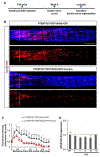
Figure 4:. DCLK1/2 are required for optic nerve regeneration induced by PTEN deletion.
(A) Experimental scheme. (B) Representative confocal images of optic nerve sections at 2 weeks post-injury from PTENf/f, or PTENf/f//DCLK1f/f/DCLK2f/f (P−/−D1−/−D2−/−) mice with AAV2-Cre injection. Scale bar: 100μm. (C) Quantification of regenerative axons from (B). ***: p<0.001 (n=5 per group). (D, E) Quantification of RGC survival (D) and phospho-S6 positive neurons (E) in the four groups. ***p<0.001 (n=5).
Figure 5:
Figure 5:. DCLK1/2 are required for sciatic nerve regeneration.
(A) Time line of the experimental procedure to study sciatic nerve regeneration. (B) Representative confocal images of the sciatic nerve sections 3 days post-injury from PTENf/fDCLK1f/f/DCLK2f/f mice with AAV8-GFP (control) or AAV8-GFP-IRES-Cre intrathecal injection. Regenerative axons are labeled with anti-SCG10 antibody (red) and sections are co-stained with anti-Tuj1 antibody (blue). White dashed line indicates the crush site. Scale bar: 500μm. (C) Quantification of regenerative axons from (B). ***: p<0.001, **: p<0.01 and *: p<0.05 (n=4-5 per group). (D) Histograms showing the expression of dclk2 by RT-qPCR on RNA extracted from whole DRG after sciatic nerve crush as indicated. dclk2 expression is normalized to GADPH.
Figure 6:
Figure 6:. Both MT-binding and S/P-rich domains of DCX can promote axon regeneration after optic nerve injury.
(A) Structures of DCX, DCLK1/2, DCX-270 and DCX-S/P. (B) Quantification of RGC survival in wild type mice infected by individual AAV2 vectors. ***: p<0.001. At least 3 animals per group. (C) Representative confocal images of optic nerve sections from PTENf/f mice with intravitreal injection of AAV2-Cre and one of the following: AAV2-PLAP, AAV2-DCX, AAV2-DCX270 or AAV2-DCX-S/P. The red stars indicate the crush site. Scale bar: 100μm. (D) Quantification of the number of regenerating axons in different groups. The number of regenerating axons seen at 0.5 mm from the crush site in the mice with DCX-270 or DCX-S/P are statistically different from those with AAV2-PLAP or AAV2-DCX. ANOVA test corrected with Bonferroni’s post-test. At least 5 animals per group. (E) Percentage of growth cone-forming neurites within the 60 min following laser cut in the explants from PTENf/f mice with intravitreal injection of AAV2-Cre and one of the following: AAV2-PLAP, AAV2-DCLK2, AAV2-DCX270 or AAV2-DCX-S/P. Chi-2 test, ***: p<0.001, *: p<0.05. The data in control and DCLK2 groups are the same as presented in Figure 3E. (F) Representative confocal images of optic nerve sections of PTENf/f mice injected with AAV2-Cre and AAV2-PLAP or PTENf/f/DCLK1f/f/DCLK2f/f mice injected with AAV2-Cre and one of the following: AAV2-DCLK2, AAV2-DCX, AAV2-DCX-270 or AAV2-DCX-S/P. The red stars indicate the crush site. Scale bar: 100μm. (G) Quantification of the number of regenerating axons in the groups shown in (D). The numbers of regenerating axons seen in the mice with DCX-270 or DCX-S/P are statistically different at all points measured from those with AAV2-PLAP or AAV2-DCX or AAV2-DCLK2. ANOVA test corrected with Bonferroni’s post-test.
Figure 7:
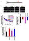
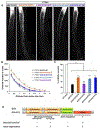
Figure 7:. DCX prevents axotomy-triggered F-actin destabilization in injured axon terminals.
(A) Experimental scheme. The red arrowhead indicates the laser cut site (100 μm away from the axonal tip). The blue line indicates the region used for quantification. (B) Micrograph of Lifeact-tdTomato fluorescence in neurites after laser cut in retinal explants from the PTENf/f mice injected with AAV2-Cre, AAV2-tdtomato-lifeact and one of the following: AAV2-PLAP, AAV2-DCLK2, AAV2-DCX-270-GFP or AAV2-DCX-SP-GFP. Time lapses began 1min post axotomy due to technical restraints of the imaging system (see Methods). Scale bar: 5μm. White arrowheads indicate laser cut site. (C) Quantification of Lifeact-tdTomato intensity from (B) during 10 min after laser cut. Lines correspond to the average of 8 samples per condition. (D) Decrease rate of F-actin intensity over time from (A). ANOVA test corrected with Bonferroni’s post-test ***p<0.001. (n=8). (E) Graph showing the quantification of phalloidin intensity after laser ablation and normalized to the precut phalloidin intensity (at least 10 axons per condition) ANOVA test corrected with Bonferroni’s post-test **p<0.01, *p<0.05.
Figure 8:
Figure 8:. The effects of DCX mutants on neuronal survival and axon regeneration.
Representative confocal images of optic nerve sections of PTENf/f mice injected with AAV2-Cre and AAV2-PLAP, AAV2-DCX-S297A, AAV2-DCX-S47R, AAV2-DCX-K174E, or AAV2-DCX-W146C. The red stars indicate the crush site. Scale bar: 100μm. Quantification of the number of regenerating axons in the groups (at least 4 animals per group) shown in (A). ANOVA test corrected with Bonferroni’s, post-test *p<0.01, ***p<0.001. The number of regenerating axons seen in the mice with DCX-S297A are statistically different at all points measured from those with AAV2-PLAP. (C) Quantification of RGC survival in wild type mice with intravitreal injection of AAV2-PLAP, AAV2-DCX-S297A, AAV2-DCX-S47R, AAV2-DCX-K174E, or AAV2-DCX-W146C. ANOVA test corrected with Bonferroni’s, post-test ***p<0.001. Error bars indicate SEM, at least 3 animals per group. (D) Model illustrating different domains and their functions associated with neuronal survival and axon regeneration. While their neuronal survival activity is relevant to the retrograde propagation activity of the MT-binding domain, their activity on axon regeneration requires both the anterograde transport activity of MT-binding domain and the F-actin-stabilizing activity of the S/P-rich actin-regulatory domain. The retrograde activity of the MT-binding domains in axon regeneration remains undetermined. The kinase domain of DCLKs is not required for these activities, but we cannot rule out a possible modulatory effect.

References

    1. Abe N, Borson SH, Gambello MJ, Wang F, and Cavalli V (2010). Mammalian target of rapamycin (mTOR) activation increases axonal growth capacity of injured peripheral nerves. J Biol Chem 285, 28034–28043. - PMC - PubMed
    1. Bielas SL, Serneo FF, Chechlacz M, Deerinck TJ, Perkins GA, Allen PB, Ellisman MH, and Gleeson JG (2007). Spinophilin facilitates dephosphorylation of doublecortin by PP1 to mediate microtubule bundling at the axonal wrist. Cell 129, 579-591. - PMC - PubMed
    1. Blackmore MG, Moore DL, Smith RP, Goldberg JL, Bixby JL, and Lemmon VP (2010). High content screening of cortical neurons identifies novel regulators of axon growth. Mol Cell Neurosci 44, 43–54. - PMC - PubMed
    1. Bradke F, Fawcett JW, and Spira ME (2012). Assembly of a new growth cone after axotomy: the precursor to axon regeneration. Nat Rev Neurosci 13, 183–193. - PubMed
    1. Campbell I (2007). Chi-squared and Fisher-Irwin tests of two-by-two tables with small sample recommendations. Stat Med 26, 3661–3675. - PubMed

Publication types

MeSH terms