Review
. 2015 Nov;53(6):1241-54.
doi: 10.1016/j.rcl.2015.06.009.

Radiological Evaluation of Bowel Ischemia

Affiliations
Review

Radiological Evaluation of Bowel Ischemia

Harpreet S Dhatt et al. Radiol Clin North Am. 2015 Nov.

Abstract

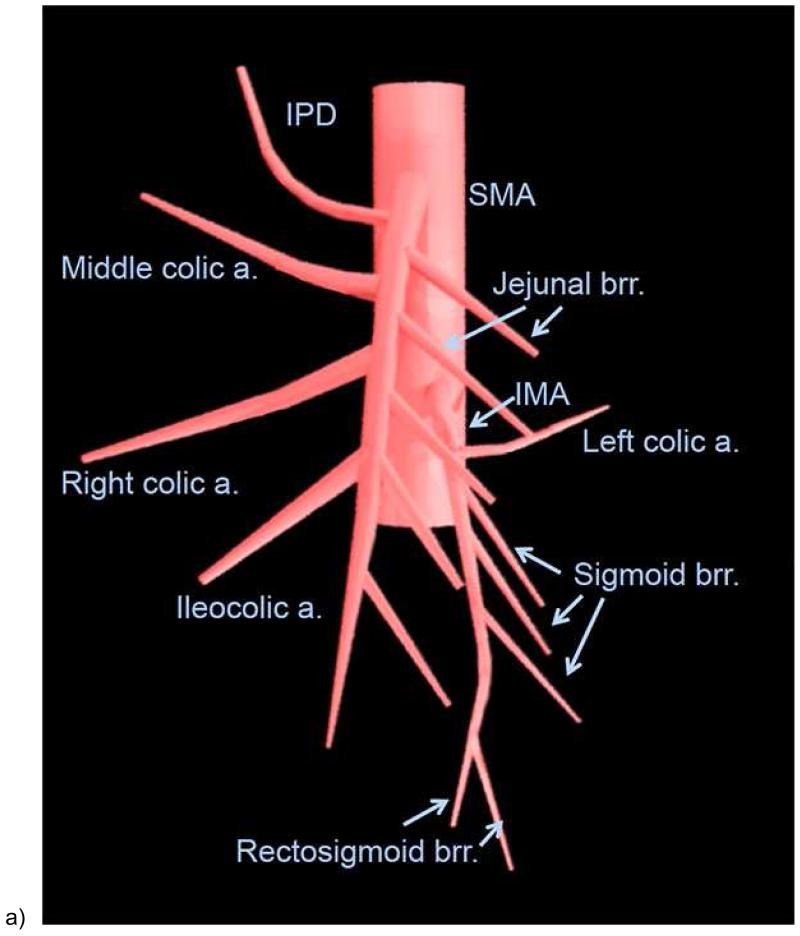
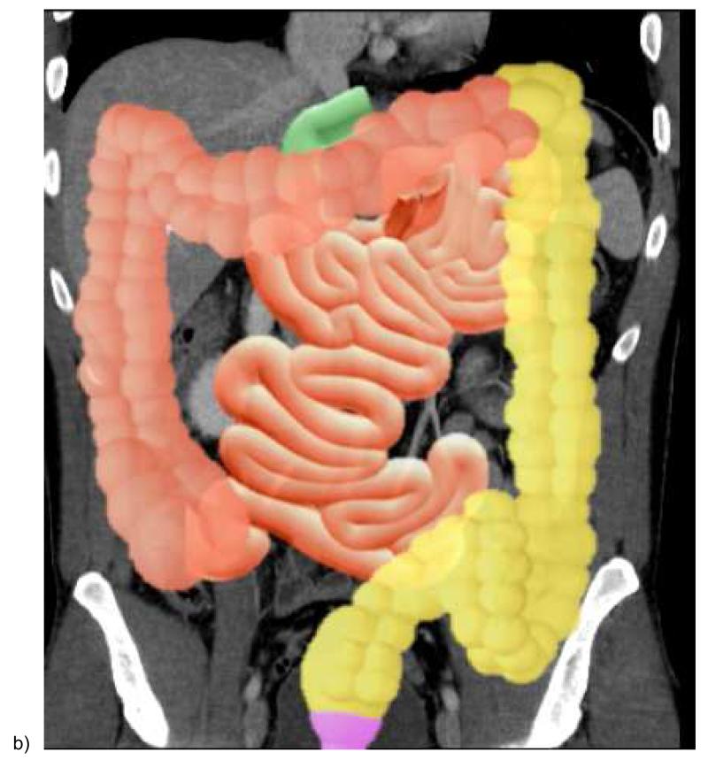
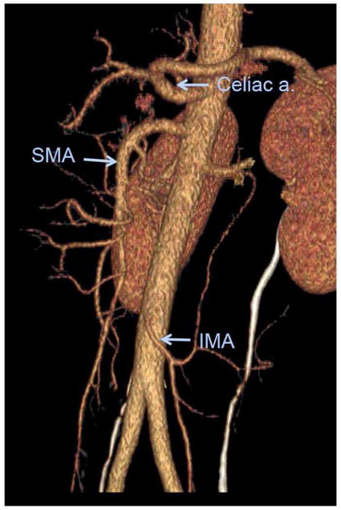
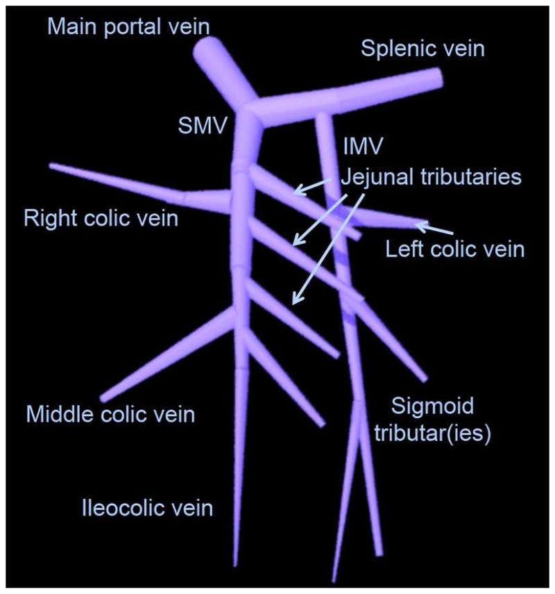
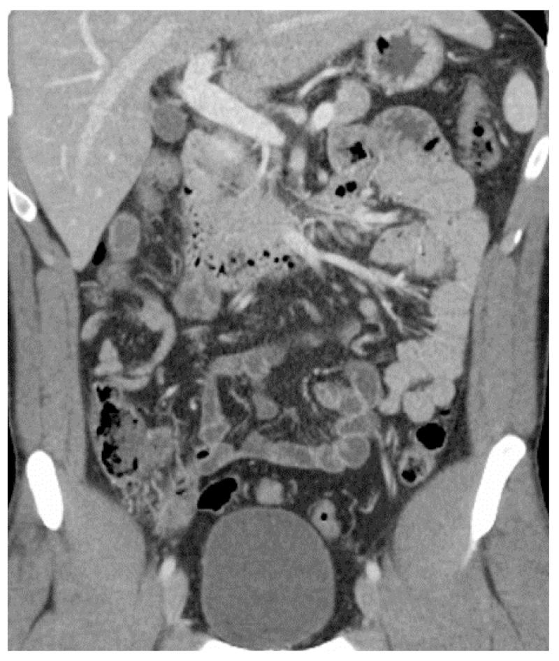
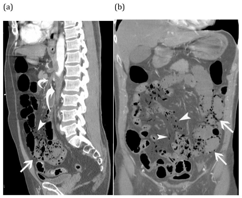
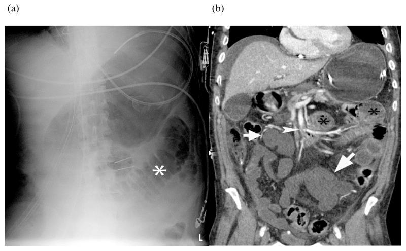
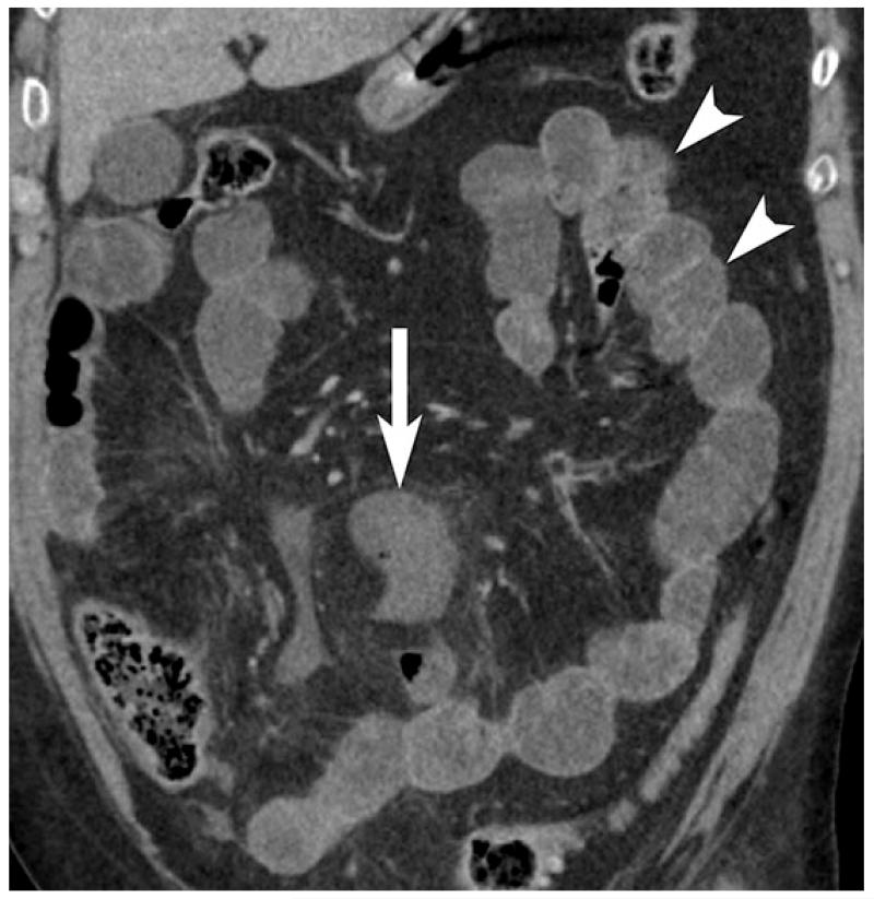
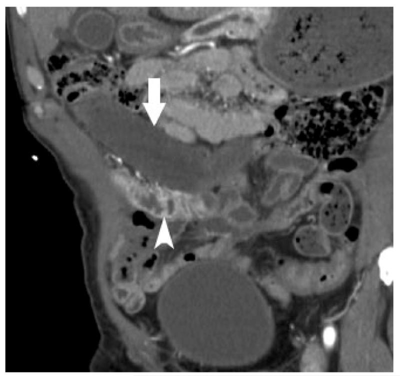
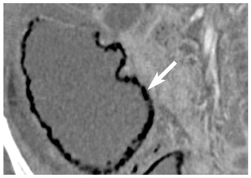
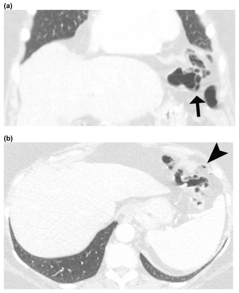
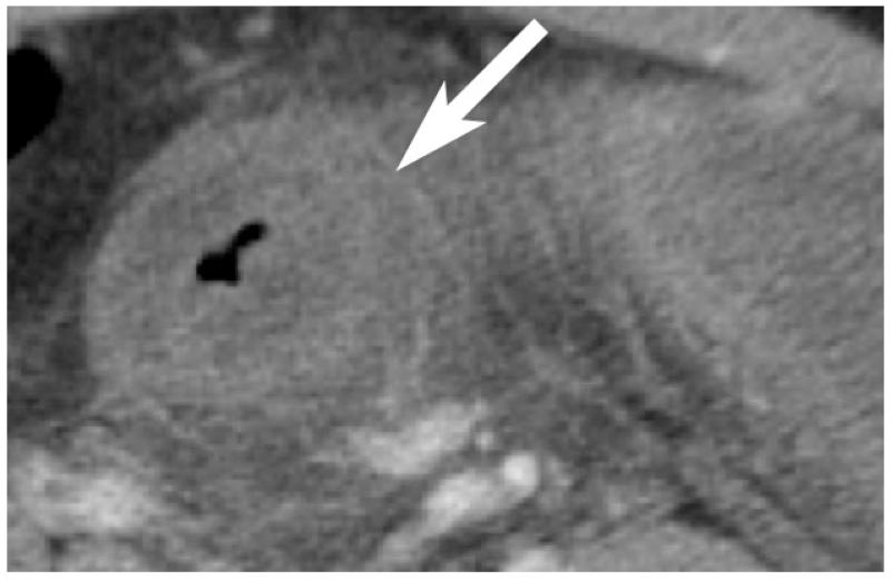
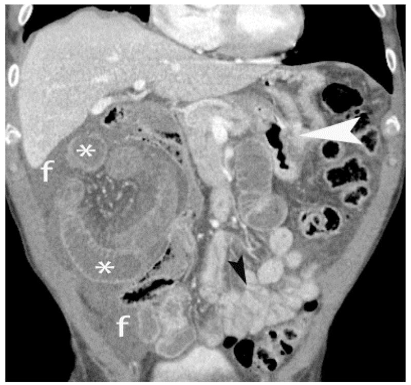
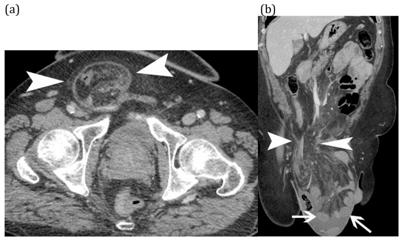
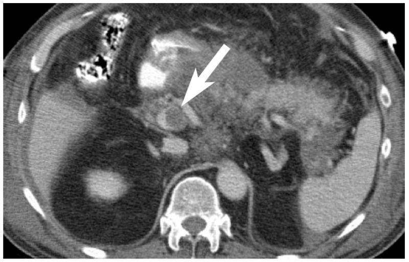
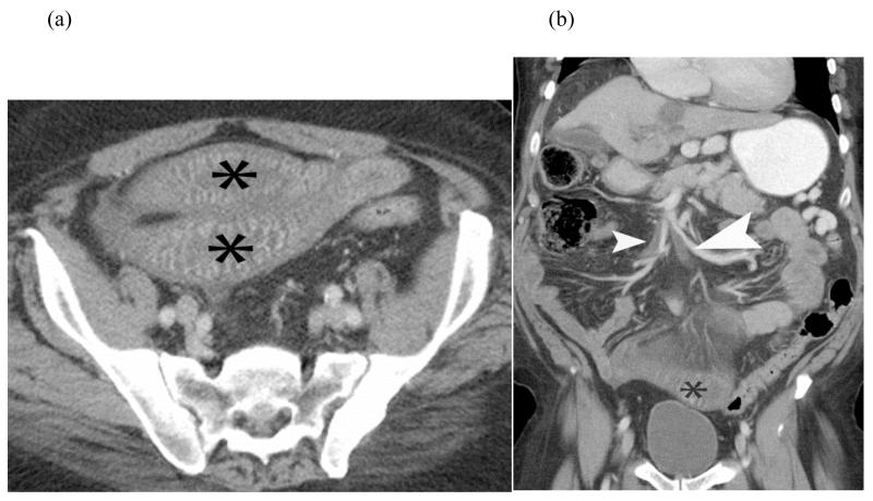
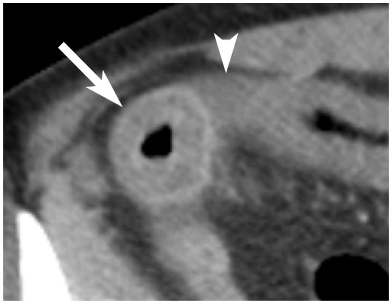
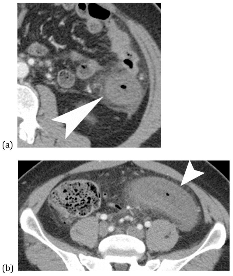
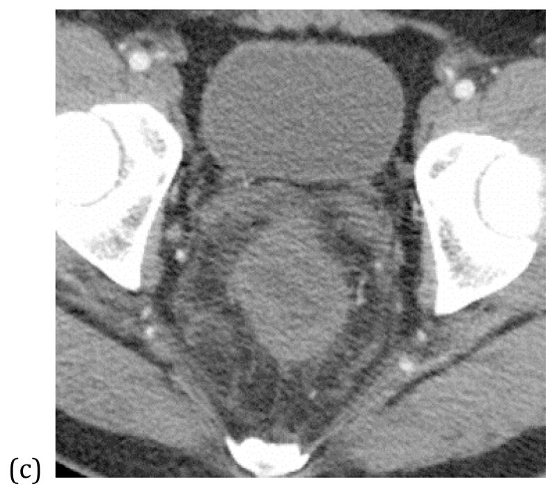
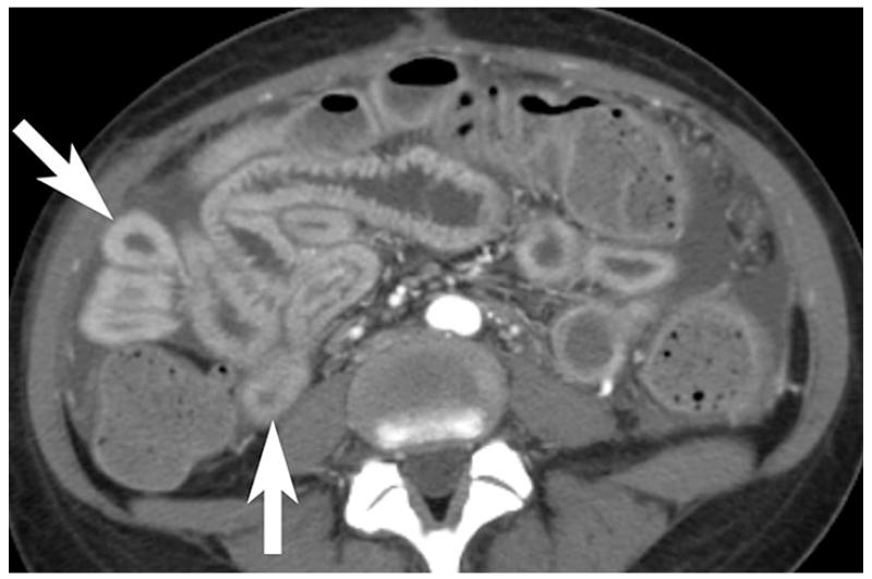
Intestinal ischemia, which refers to insufficient blood flow to the bowel, is a potentially catastrophic entity that may require emergent intervention or surgery in the acute setting. Although the clinical signs and symptoms of intestinal ischemia are nonspecific, computed tomography (CT) findings can be highly suggestive in the correct clinical setting. In our article, we review the CT diagnosis of arterial, venous, and nonocclusive intestinal ischemia. We discuss the vascular anatomy, pathophysiology of intestinal ischemia, CT techniques for optimal imaging, key and ancillary radiological findings, and differential diagnosis.

Keywords: Bowel infarction; Bowel ischemia; Computed tomography; Intestinal ischemia; Mesenteric artery occlusion; Mesenteric ischemia; Oral contrast; Pneumatosis intestinalis.


Figures

Figure 1
Figure 1
Illustration of the mesenteric arteries (a) and bowel segments supplied by mesenteric arteries (b). SMA = superior mesenteric artery. IPD = inferior pancreaticoduodenal artery. a.= artery; brr. = branch artery; IMA = inferior mesenteric artery. The duodenum, jejunum, ileum, and colon proximal to the splenic flexure is supplied by the superior mesenteric artery (bowel with orange color), and the descending and sigmoid colon and upper rectum are supplied by the inferior mesenteric artery (bowel in yellow color). The distal most rectum is supplied by the middle and inferior rectal arteries from the internal iliac artery (bowel in purple color)
Figure 1
Figure 1
Illustration of the mesenteric arteries (a) and bowel segments supplied by mesenteric arteries (b). SMA = superior mesenteric artery. IPD = inferior pancreaticoduodenal artery. a.= artery; brr. = branch artery; IMA = inferior mesenteric artery. The duodenum, jejunum, ileum, and colon proximal to the splenic flexure is supplied by the superior mesenteric artery (bowel with orange color), and the descending and sigmoid colon and upper rectum are supplied by the inferior mesenteric artery (bowel in yellow color). The distal most rectum is supplied by the middle and inferior rectal arteries from the internal iliac artery (bowel in purple color)
Figure 2
Figure 2
Volume rendered oblique sagittal reformation of normal CT angiogram shows the major mesenteric arteries.
Figure 3
Figure 3
Illustration of normal major mesenteric veins. SMV = superior mesenteric vein. IMV = inferior mesenteric vein.
Figure 4
Figure 4
Normal small bowel on CT - Coronal image of normal small bowel. Notice the relative increased enhancement of the jejunum in the left upper quadrant compared to the ileum in the right lower quadrant. The apparent jejunal hyperenhancement is due to the higher fold density of the valvulae conniventes in collapsed jejunum than ileum.
Figure 5
Figure 5
Arterial ischemia from SMA thrombosis – Elderly man presented with three days of abdominal pain and anorexia. Sagittal (a) and coronal (b) CT images show pneumatosis intestinalis (white arrows) and mesenteric venous gas (white arrowheads) associated with extensive clot in the aorta (large curved arrow) extending into the celiac trunk and SMA (small curved arrow).
FIGURE 6
FIGURE 6
Plain radiograph of small bowel ischemia – Plain radiograph (a) shows a focally dilated “paper thin” segment of small bowel (*) that had persisted over several consecutive examinations. Subsequent coronal reformatted CT image (b) of the same patient shows the corresponding dilated segment (*) as well as other fluid filled segments of small bowel with absent mural enhancement (white arrow) and clot in the SMA (white arrow head). At laparotomy, 220 centimeters of dead bowel was found.
Figure 7
Figure 7
54 year old with SMA thrombus causing arterial bowel ischemia. Thin hypoenhancing bowel wall (arrow) and associated subtle mesenteric fat stranding is seen on coronal contrast enhanced CT image. Mural enhancement of non-ischemic jejunum (arrowheads) is seen in the left upper abdomen.
Figure 8
Figure 8
Coronal contrast enhanced CT shows pale arterial ischemia with absent mural enhancement in a segment of small bowel (arrow). An adjacent segment of small bowel shows mural hyperenhancement (arrowhead), indicating bowel reperfusion injury.
Figure 9
Figure 9
Coronal CT showing pneumatosis intestinalis; gas within the bowel wall (white arrow) can be suggestive bowel ischemia and infarction in the appropriate clinical setting. In the absence of other concerning CT findings, clinical signs, or symptoms of bowel ischemia, a benign cause pneumatosis should be considered.
Figure 10
Figure 10
Benign pneumatosis – CT images in lung window of a patient who presented with abdominal pain after trauma. Coronal image (a) shows pneumatosis cystoides coli (black arrow), while the axial images (b) shows small volume of pneumoperitoneum (black arrow head). Patient was admitted for observation and discharged without any intervention as his pain resolved spontaneously.
Figure 11
Figure 11
Abnormally thickened bowel (white arrow) with mesenteric fat stranding and slightly decreased mural enhancement is non-specific; etiologies include bowel ischemia, edema, intramural hemorrhage and/or superimposed infection.
Figure 12
Figure 12
Closed loop obstructionleading to mesenteric ischemia– CT scan of a patient who presented with sudden abdomen pain and nausea. A focally dilated segment of ischemic small bowel (*) with collapsed proximal (white arrowhead) and distal (black arrow head) small bowel is seen. The dilated ischemic bowel shows less enhancement than the collapsed segments of normal small bowel. The presence of adjacent free fluid (f) and mesenteric edema is also concerning for early ischemia.
Figure 13
Figure 13
Strangulated small bowel – Axial (a) and coronal (b) CT images of incarcerated small bowel (white arrows) in a large right inguinal hernia (white arrow heads). CT findings include hypoenhancement of the small bowel wall (arrows) with adjacent fluid and fat stranding of the associated mesentery in the hernia sac.
Figure 14
Figure 14
Contrast enhanced CT shows low-attenuation clot within SMV (arrow) in a patient with pancreatitis
Figure 15
Figure 15
Venous bowel ischemia from SMV thrombosis - Axial (a) and coronal reformatted (b) CT images of a patient with history of cirrhosis presenting with abdominal pain. Axial CT image shows marked bowel wall thickening with hyperenhancement and mesenteric edema. Coronal image shows thrombosis of the SMV. Venous ischemia presents with marked bowel thickening and may show some bowel wall enhancement, unlike arterial ischemia which often shows normal to thinned wall thickness and absent mural enhancement.
Figure 16
Figure 16
“Target-appearance of bowel”; venous occlusion resulting in bowel edema with hyperenhancement (arrow) of serosal/subserosal layers, mesenteric stranding, and small adjacent free fluid (arrowhead).
Figure 17
Figure 17
Veno-occlusive disease – Three axial CT images (a-c) show marked bowel wall thickening with poor enhancement of the colon (white arrowhead) continuously from the descending colon to the rectum. Associated mesenteric fat stranding is seen.
Figure 17
Figure 17
Veno-occlusive disease – Three axial CT images (a-c) show marked bowel wall thickening with poor enhancement of the colon (white arrowhead) continuously from the descending colon to the rectum. Associated mesenteric fat stranding is seen.
Figure 18
Figure 18
Shock bowel; mucosal hyperenhancement of thick walled small bowel (arrows) and ascites suggests recent hypotension.
FIGURE 19 (a) and (b)
Shock bowel from hypotension – Coronal CT images of an elderly man with presented sepsis and hypotension. The first image (a) shows non-enhancement of a small bowel segment (*) compatible with small bowel ischemia. Additional images (b) shows evidence of global hypotension and shock with renal cortical necrosis (black arrow heads) and splenic infarcts (white arrows).
FIGURE 20
FIGURE 20
Watershed colonic Ischemia - Axial (a) and sagittal (b) CT images of a patient with hypotension shows segmental bowel wall thickening and poor mural enhancement of the descending colon (white arrowhead) with sparing of the transverse colon (white arrow).

References

    1. Cartwright SL, Knudson MP. Evaluation of acute abdominal pain in adults. American family physician. 2008;77(7):971–8. - PubMed
    1. Ruotolo RA, Evans SR. Mesenteric ischemia in the elderly. Clinics in geriatric medicine. 1999;15(3):527–57. - PubMed
    1. Kumar S, Sarr MG, Kamath PS. Mesenteric venous thrombosis. The New England journal of medicine. 2001;345(23):1683–8. - PubMed
    1. Stamatakos M, Stefanaki C, Mastrokalos D, et al. Mesenteric ischemia: still a deadly puzzle for the medical community. The Tohoku journal of experimental medicine. 2008;216(3):197–204. - PubMed
    1. Huang HH, Chang YC, Yen DH, et al. Clinical factors and outcomes in patients with acute mesenteric ischemia in the emergency department. Journal of the Chinese Medical Association: JCMA. 2005;68(7):299–306. - PubMed

MeSH terms