. 2015 Nov 11;18(5):582-92.
doi: 10.1016/j.chom.2015.10.007. Epub 2015 Oct 29.

Normalization of Host Intestinal Mucus Layers Requires Long-Term Microbial Colonization

Affiliations

Normalization of Host Intestinal Mucus Layers Requires Long-Term Microbial Colonization

Malin E V Johansson et al. Cell Host Microbe. .

Abstract

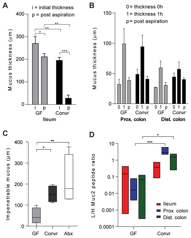
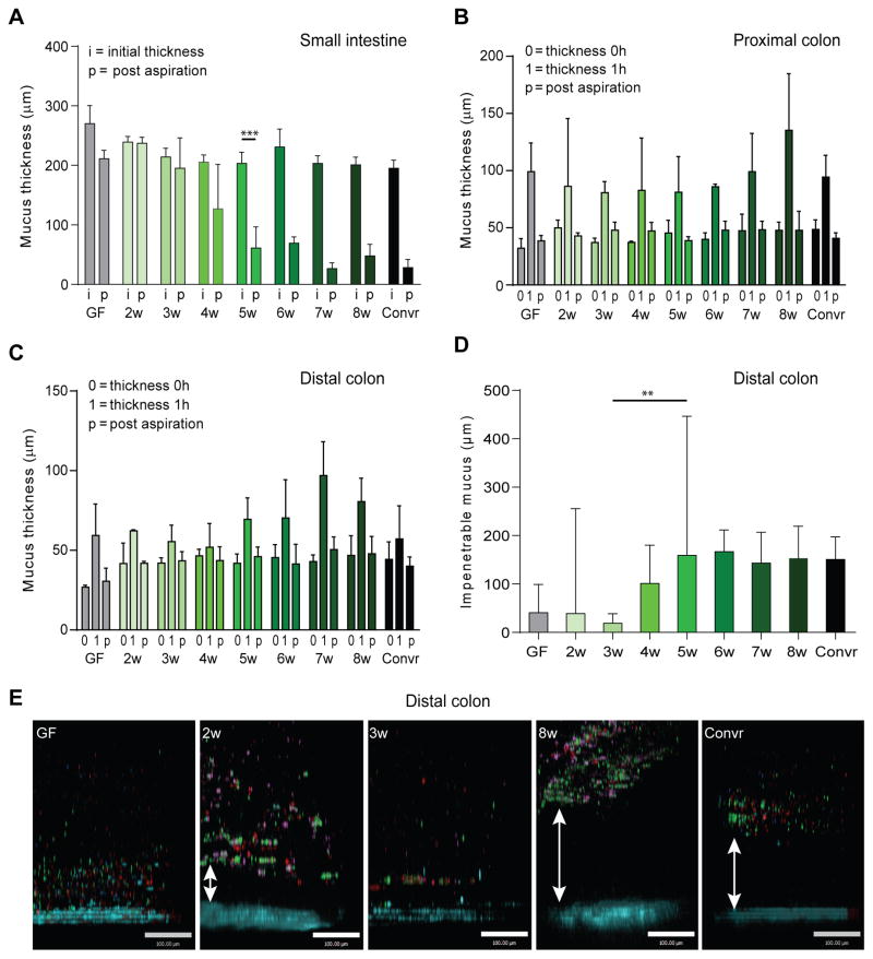
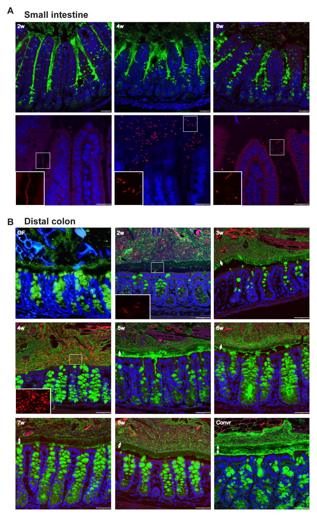
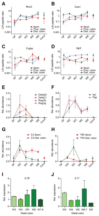
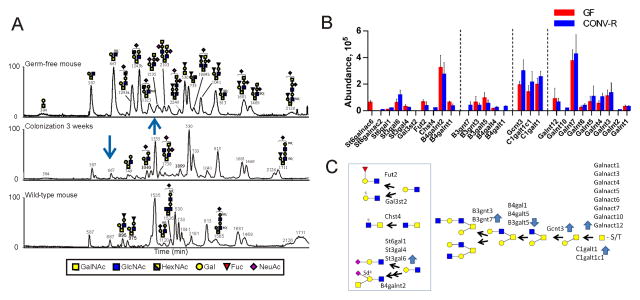
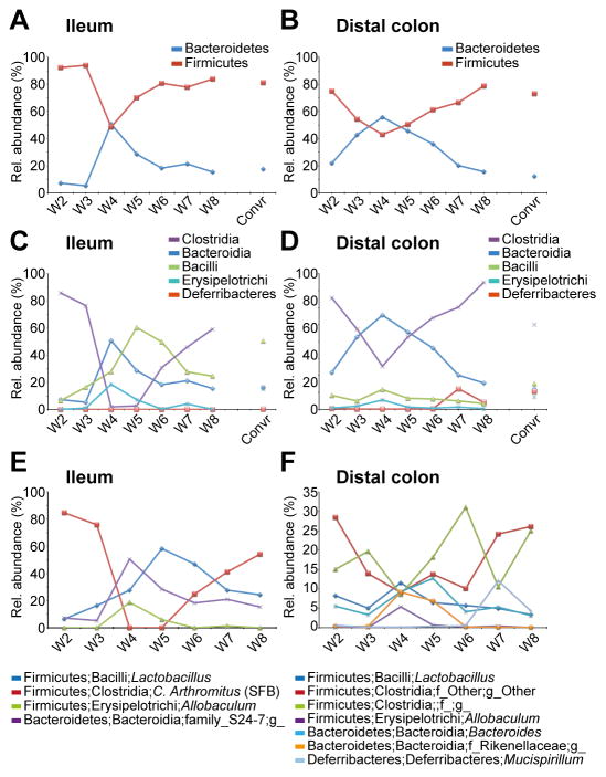
The intestinal mucus layer provides a barrier limiting bacterial contact with the underlying epithelium. Mucus structure is shaped by intestinal location and the microbiota. To understand how commensals modulate gut mucus, we examined mucus properties under germ-free (GF) conditions and during microbial colonization. Although the colon mucus organization of GF mice was similar to that of conventionally raised (Convr) mice, the GF inner mucus layer was penetrable to bacteria-sized beads. During colonization, in which GF mice were gavaged with Convr microbiota, the small intestine mucus required 5 weeks to be normally detached and colonic inner mucus 6 weeks to become impenetrable. The composition of the small intestinal microbiota during colonization was similar to Convr donors until 3 weeks, when Bacteroides increased, Firmicutes decreased, and segmented filamentous bacteria became undetectable. These findings highlight the dynamics of mucus layer development and indicate that studies of mature microbe-mucus interactions should be conducted weeks after colonization.


Figures

Figure 1
Figure 1. Mucus in germ free and conventionalized mice
(A) Mucus thickness in distal small intestine of germ free (GF) or conventionally raised (Convr) mice was measured as initial thickness (i) or post-aspiration (p). The mucus thickness was significantly different before and post-aspiration. The mucus remaining after aspiration was significantly different between GF and Convr mice revealing attached mucus in GF mice. (B) Mucus thickness measured in distal colon during 1h. The results are reported as the thickness of the initially attached mucus (0), mucus thickness after 1h (1) and the attached mucus after 1h post aspiration (p). No differences between GF animals and Convr controls were observed. (C) Mucus penetrability to beads the size of bacteria (0.5–2 μm) was measured by adding beads to newly secreted mucus and allowing sedimentation for 40 min followed by collecting confocal Z-stack images. The impenetrable mucus fraction was analyzed in distal colon and revealed significant differences between GF and Convr mice. Treatment with antibiotics for 3 weeks did not impair the mucus penetrability (Abx). (D) Muc2 protein amounts were analyzed by mass spectrometry with heavy labelled peptides and significantly higher amounts of Muc2 were observed in colon of Convr mice compare to GF (*= P<0.05, **= P<0.01, ***= P<0.001, ****= P<0.0001).
Figure 2
Figure 2. Mucus during colonization with a complex microbiota
(A) Mucus thickness in distal small intestine was measured as in Fig. 1A during 2 to 8 weeks of colonization. The mucus was removable after 4 weeks of colonization. (B–C) The mucus thickness was measured over 1 h in proximal (B) and distal (C) colon as in Fig. 1B during 2–8 weeks of colonization. (D) Mucus penetrability was analyzed in distal colon during colonization as in Fig. 1C. An impenetrable mucus developed after 6 weeks. (E) Representative pictures of confocal Z-stacks from the penetrability experiments with beads (0.5 μm red, 1 μm purple, 2 μm green) close to the epithelium (blue) in GF, 2 and 3-week colonized mice. Mucus separated the beads from the epithelium in Convr mice and after 8 weeks of conventionalization. The scale bar represents 100 μm. **, P=0.016, ***, P=0.002.
Figure 3
Figure 3. Immunohistology of small intestine and colon during conventionalization
Immunostaining of sections from distal small intestine (A) and distal colon (B) of Muc2 (green) with FISH detecting bacteria (red) and Hoechst counterstain of DNA (blue). Double arrows indicate the inner mucus layer. Inserts show the bacteria magnified in the boxed areas. Typically, the middle part of the inner mucus layer is not stained as well as Muc2 at other locations. Scale bars in A, upper panel and B, 50 μm; A, lower panel, 25 μm.
Figure 4
Figure 4. Alterations in mucus proteins after colonization of germ free mice
Amounts of the mucus protein Muc2 (A), Clca1 (B), Fcgbp (C), and Agr2 (D) relative to supplemented labelled internal standard peptides in the ileum, proximal colon and distal colon as determined by proteomics of aspirated mucus from explants. (E–F) Alterations in relative amounts of antibacterial peptides/proteins (E), immunoglobulin J chain (IgJ) and the polymeric immunoglobulin receptor (Pigr) (F) in ileum as determined by proteomics of aspirated mucus from explants. (G–H) Alterations of the protein amounts of complement factor 3 (C3) and trefoil factor 3 (Tff3) in ileum and distal colon as determined by proteomics of aspirated mucus from explants. (I–J) mRNA levels of IL1β (I) and IL17 (J) as determined by Q-PCR in whole distal colon tissue. See also Fig. S1, S5, and Table S2.
Figure 5
Figure 5. Glycosylation alterations during colonization
(A) Base peak chromatograms of the capillaryLC-MS/MS analyses of the mouse midcolon Muc2 O-glycans of a GF mouse (top), GF conventionalized for 3 weeks (middle), and Convr WT mouse (bottom). The most abundant glycans in the LC profiles are annotated in the figure. Arrows point to major alterations. (B) Relative abundance of glycosyltransferases in the epithelial cells of GF and Convr mice as determined by proteomic analyses. (C) Proposed biosynthetic pathways and corresponding glycosyltransferases for the observed Muc2 glycans. Blue arrows show increased or decreased levels of the transferases upon colonization. See also Table S3–S4.
Figure 6
Figure 6. Microbiota alterations in ileum and distal colon mucus during colonization
(A–B) The relative abundance (%) of the Firmicutes and Bacteroidetes phyla in ileum mucus (A) and distal colon mucus (B) at weeks 2–8 after colonization. (C–D) The relative abundance (%) of classes found in ileum mucus (C) and distal colon mucus (D) at weeks 2–8 after colonization. (E–F) The relative abundance (%) of selected, most abundant, genera in ileum mucus (E) and distal colon mucus (F) at weeks 2–8 after colonization. n=2–3 mice/time point. For details regarding bacteria and mice, see also Figs. S2–S4, S6, Table S5.

References

    1. Backhed F, Ley RE, Sonnenburg JL, Peterson DA, Gordon JI. Host-Bacterial Mutualism in the Human Intestine. Science. 2005;307:1915–1920. - PubMed
    1. Backhed F, Roswall J, Peng Y, Feng Q, Jia H, Kovatcheva-Datchary P, Li Y, Xia Y, Xie H, Zhong H, et al. Dynamics and Stabilization of the Human Gut Microbiome during the First Year of Life. Cell Host Microbe. 2015;17:690–703. - PubMed
    1. Belzer C, Gerber GK, Roeselers G, Delaney M, DuBois A, Liu Q, Belavusava V, Yeliseyev V, Houseman A, Onderdonk A, et al. Dynamics of the Microbiota in Response to Host Infection. PLoS ONE. 2014;9:e95534. - PMC - PubMed
    1. Booijink CC, El-Aidy S, Rajilic-Stojanovic M, Heilig HG, Troost FJ, Smidt H, Kleerebezem M, de Vos WM, Zoetendal EG. High temporal and inter-individual variation detected in the human ileal microbiota. Environ Microbiol. 2010;12:3213–3227. - PubMed
    1. Bry L, Falk PG, Midtvedt T, Gordon JI. A model of host-microbial interactions in an open mammalian ecosystem. Science. 1996;273:1380–1383. - PubMed

Publication types