Cerebral Perfusion and Gray Matter Changes Associated With Chemotherapy-Induced Peripheral Neuropathy
- PMID: 26527786
- PMCID: PMC4822503
- DOI: 10.1200/JCO.2015.62.1276
Cerebral Perfusion and Gray Matter Changes Associated With Chemotherapy-Induced Peripheral Neuropathy
Abstract
Purpose: To investigate the longitudinal relationship between chemotherapy-induced peripheral neuropathy (CIPN) symptoms (sx) and brain perfusion changes in patients with breast cancer. Interaction of CIPN-sx perfusion effects with known chemotherapy-associated gray matter density decrease was also assessed to elucidate the relationship between CIPN and previously reported cancer treatment-related brain structural changes.
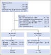
Methods: Patients with breast cancer treated with (n = 24) or without (n = 23) chemotherapy underwent clinical examination and brain magnetic resonance imaging at the following three time points: before treatment (baseline), 1 month after treatment completion, and 1 year after the 1-month assessment. CIPN-sx were evaluated with the self-reported Functional Assessment of Cancer Therapy/Gynecologic Oncology Group-Neurotoxicity four-item sensory-specific scale. Perfusion and gray matter density were assessed using voxel-based pulsed arterial spin labeling and morphometric analyses and tested for association with CIPN-sx in the patients who received chemotherapy.
Results: Patients who received chemotherapy reported significantly increased CIPN-sx from baseline to 1 month, with partial recovery by 1 year (P < .001). CIPN-sx increase from baseline to 1 month was significantly greater for patients who received chemotherapy compared with those who did not (P = .001). At 1 month, neuroimaging showed that for the group that received chemotherapy, CIPN-sx were positively associated with cerebral perfusion in the right superior frontal gyrus and cingulate gyrus, regions associated with pain processing (P < .001). Longitudinal magnetic resonance imaging analysis in the group receiving chemotherapy indicated that CIPN-sx and associated perfusion changes from baseline to 1 month were also positively correlated with gray matter density change (P < .005).
Conclusion: Peripheral neuropathy symptoms after systemic chemotherapy for breast cancer are associated with changes in cerebral perfusion and gray matter. The specific mechanisms warrant further investigation given the potential diagnostic and therapeutic implications.
© 2015 by American Society of Clinical Oncology.
Conflict of interest statement
Authors' disclosures of potential conflicts of interest are found in the article online at
Figures


Comment in
-
Painful Hands and Feet After Cancer Treatment: Inflammation Affecting the Mind-Body Connection.J Clin Oncol. 2016 Mar 1;34(7):649-52. doi: 10.1200/JCO.2015.64.7479. Epub 2015 Dec 23. J Clin Oncol. 2016. PMID: 26700128 No abstract available.
References
-
- Wickham R. Chemotherapy-induced peripheral neuropathy: A review and implications for oncology nursing practice. Clin J Oncol Nurs. 2007;11:361–376. - PubMed
-
- Seretny M, Currie GL, Sena ES, et al. Incidence, prevalence, and predictors of chemotherapy-induced peripheral neuropathy: A systematic review and meta-analysis. Pain. 2014;155:2461–2470. - PubMed
-
- Argyriou AA, Bruna J, Marmiroli P, et al. Chemotherapy-induced peripheral neuropathy (CIPN): An update. Crit Rev Oncol Hematol. 2012;82:51–77. - PubMed
Publication types
MeSH terms
Substances
Grants and funding
- R01 AG019771/AG/NIA NIH HHS/United States
- S10 RR027710/RR/NCRR NIH HHS/United States
- UL1 RR025761/RR/NCRR NIH HHS/United States
- RR020128/RR/NCRR NIH HHS/United States
- P30 AG10133/AG/NIA NIH HHS/United States
- P30 AG010133/AG/NIA NIH HHS/United States
- R25 CA117865/CA/NCI NIH HHS/United States
- U54 HD062484/HD/NICHD NIH HHS/United States
- R01 CA082709/CA/NCI NIH HHS/United States
- T32 CA117865/CA/NCI NIH HHS/United States
- R01 HD062484/HD/NICHD NIH HHS/United States
- P30 CA082709/CA/NCI NIH HHS/United States
- UL1 TR001108/TR/NCATS NIH HHS/United States
- C06 RR020128/RR/NCRR NIH HHS/United States
- RR027710-01/RR/NCRR NIH HHS/United States
- R01 AG19771/AG/NIA NIH HHS/United States
- R01 CA101318/CA/NCI NIH HHS/United States
LinkOut - more resources
Full Text Sources
Other Literature Sources
Medical

