

A rare variation of the digastric muscle
- PMID: 26527971
- PMCID: PMC4462457
A rare variation of the digastric muscle
Abstract
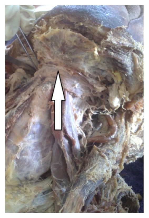
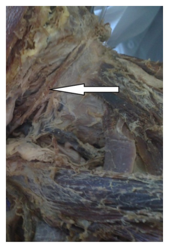
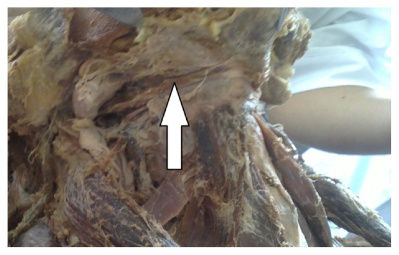
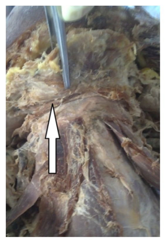
The digastric muscle is composed by two muscle bellies: an anterior and a posterior, joined by an intermediate tendon. This muscle is situated in the anterior region of the neck. The region between the hyoid bone and the mandible is divided by an anterior belly into two triangles: the submandibular situated laterally and the submental triangle which is located medially. We found that the anatomical variations described in the literature relate mainly to the anterior belly and consist of differences in shape and attachment of the muscle. During routine dissection in February 2013 in the section hall of the Department of Anatomy and Histology in Medical University - Sofia we came across a very interesting variation of the digastric muscle. The digastric muscles that presented anatomical variations were photographed using a Sony Cyber-shot DSC-T1 camera, with a Carl Zeiss Vario-Tessar lens. We found out bilateral variation of the digastric muscle in one cadaver. The anterior bellies were very thin and insert to the hyoid bone. Two anterior bellies connect each other and thus they formed a loop. The anatomical variations observed of our study related only to the anterior belly, as previously described by other authors. It is very important to consider the occurrence of the above mentioned variations in the digastric muscle when surgical procedures are performed on the anterior region of the neck.
Keywords: digastric muscle; hyoid bone; neck muscles; variations.
Figures
References
-
- Andreo JC, Navarro JAC, Toledo Filho JL. Anatomical study on the variations of the anterior belly of the digastric uscle. Rev Chil Anat. 1997;15(1):111–114.
-
- Bergman RA, Afifi AK, Miyauchi R. Digastricus. Illustrated Encyclopedia of Human Anatomic Variation. Opus I: Muscular system. 2002. Available from: http://www.anatomyatlases.org.
-
- Drake RL, Vogl W, Mitchell AWM. Cabeça e pescoço. In: Drake RL, Vogl W, Mitchell AWM, editors. Gray’s Anatomia para estudantes. 1ª ed. Rio de Janeiro: Elsevier; 2005. pp. 905–907.
-
- Fujimura A, Onodera M, Feng X, et al. Abnormal anterior belly of the digastric muscle: A proposal for the classification of abnormalities. Anat Science Inter. 2003;78:185–188. - PubMed
-
- Liquidato BM, Barros MD, Alves AL, Pereira CSB. Anatomical study of the digastric muscle: Variations in the anterior belly. Int J Morphol. 2007;25(4):797–800.
LinkOut - more resources
Full Text Sources