

Patterns of Change During the Dermoscopic Follow-Up of Melanocytic Lesions in High Risk Patients
- PMID: 26528046
- PMCID: PMC4508616
- DOI: 10.15386/cjmed-394
Patterns of Change During the Dermoscopic Follow-Up of Melanocytic Lesions in High Risk Patients
Abstract
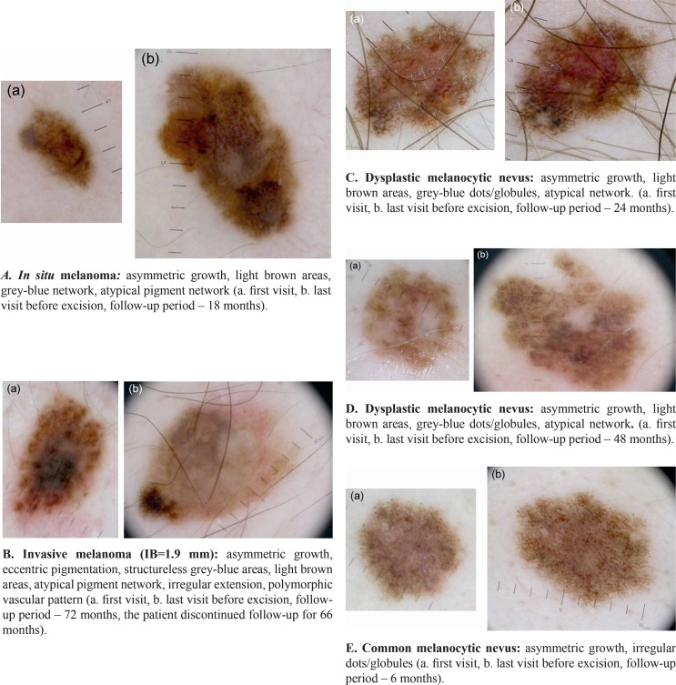
Background: Melanomas and melanocytic nevi that change over time display different change patterns, correlated with histopathological features.
Methods: We performed a retrospective analysis of the dermoscopic images corresponding to 86 lesions excised due to the changes occurred during the follow-up period in patients at high risk for melanoma, and we drew a comparison between the changes occurring in melanomas and those occurring in melanocytic nevi.
Results: There were significant differences between the models of dermoscopic change characteristic to melanoma and those characteristic to melanocytic nevi. We observed changes with high specificity for the diagnosis of melanoma - asymmetric growth (Sp=90%), new structureless grey-blue areas (Sp=97.5%) or new grey-blue network (Sp=96.25%), new pseudopods or radial streaks (Sp=95%).
Conclusion: Our study highlights highly specific changes whose presence should raise the suspicion of melanoma and lead to the excision of the lesion.
Keywords: dermoscopy; melanocytic nevi; melanoma.
Figures
Similar articles
-
Clinical and Histopathologic Characteristics of Melanocytic Lesions on the Volar Skin Without Typical Dermoscopic Patterns.JAMA Dermatol. 2019 May 1;155(5):578-584. doi: 10.1001/jamadermatol.2018.5926. JAMA Dermatol. 2019. PMID: 30865233 Free PMC article.
-
Diagnostic Performance of Dermoscopy for Distinguishing Early Melanomas and Intermediate Melanocytic Lesions From Low-Grade Dysplastic Nevi.J Cutan Med Surg. 2025 Mar 12:12034754251325508. doi: 10.1177/12034754251325508. Online ahead of print. J Cutan Med Surg. 2025. PMID: 40072505
-
Dermoscopic predictors of melanoma in small diameter melanocytic lesions (mini-melanoma): a retrospective multicentric study of 269 cases.Int J Dermatol. 2023 Aug;62(8):1040-1049. doi: 10.1111/ijd.16710. Epub 2023 May 20. Int J Dermatol. 2023. PMID: 37208996
-
Dermoscopy for the pediatric dermatologist part III: dermoscopy of melanocytic lesions.Pediatr Dermatol. 2013 May-Jun;30(3):281-93. doi: 10.1111/pde.12041. Epub 2012 Dec 18. Pediatr Dermatol. 2013. PMID: 23252411 Review.
-
Role of In Vivo Reflectance Confocal Microscopy in the Analysis of Melanocytic Lesions.Acta Dermatovenerol Croat. 2018 Apr;26(1):64-67. Acta Dermatovenerol Croat. 2018. PMID: 29782304 Review.
References
-
- Siegel R, Ward E, Brawley O, Jemal A. Cancer statistic 2011: the impact of eliminating socioeconomic and racial disparities on premature cancer deaths. CA Cancer J Clin. 2011;61:212–236. - PubMed
-
- Cockerell CJ. The pathology of melanoma. Dermatol Clin. 2012;30:445–468. - PubMed
-
- Snels DG, Hille ET, Gruis NA, Bergman W. Risk of cutaneous malignant melanoma in patients with nonfamilial atypical nevi from a pigmented lesions clinic. J Am Acad Dermatol. 1999;40:686–693. - PubMed
-
- Gandini S, Sera F, Cattaruzza MS, Pasquini P, Abeni D, Boyle P, et al. Meta-analysis of risk factors for cutaneous melanoma: I. Common and atypical naevi. Eur J Cancer. 2005;41(1):28–44. - PubMed
-
- Bauer J, Blum A, Strohhacker U, Garbe C. Surveillance of patients at high risk for cutaneous malignant melanoma using digital dermoscopy. Br J Dermatol. 2005;152:87–92. - PubMed
LinkOut - more resources
Full Text Sources
Other Literature Sources