. 2015 Oct 15:3:63.
doi: 10.3389/fcell.2015.00063. eCollection 2015.

Uncovering the dual role of RHAMM as an HA receptor and a regulator of CD44 expression in RHAMM-expressing mesenchymal progenitor cells

Affiliations

Uncovering the dual role of RHAMM as an HA receptor and a regulator of CD44 expression in RHAMM-expressing mesenchymal progenitor cells

Mandana Veiseh et al. Front Cell Dev Biol. .

Abstract

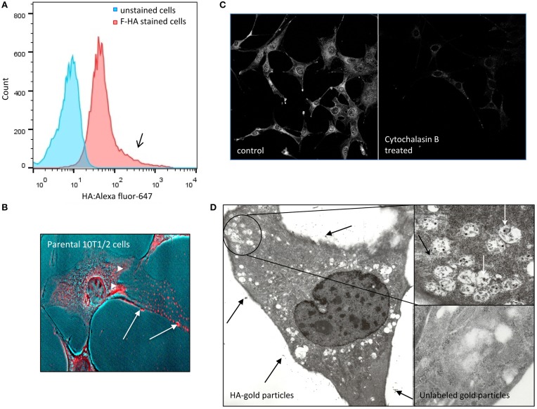
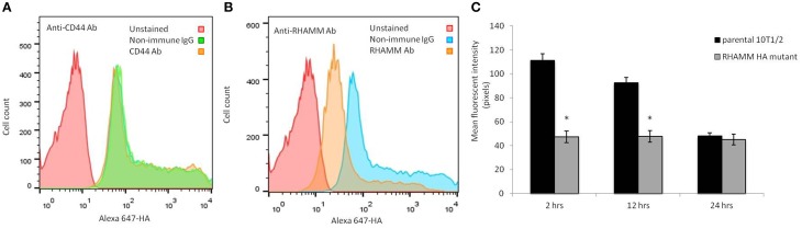
The interaction of hyaluronan (HA) with mesenchymal progenitor cells impacts trafficking and fate after tissue colonization during wound repair and these events contribute to diseases such as cancer. How this interaction occurs is poorly understood. Using 10T½ cells as a mesenchymal progenitor model and fluorescent (F-HA) or gold-labeled HA (G-HA) polymers, we studied the role of two HA receptors, RHAMM and CD44, in HA binding and uptake in non-adherent and adherent mesenchymal progenitor (10T½) cells to mimic aspects of cell trafficking and tissue colonization. We show that fluorescent labeled HA (F-HA) binding/uptake was high in non-adherent cells but dropped over time as cells became increasingly adherent. Non-adherent cells displayed both CD44 and RHAMM but only function-blocking anti-RHAMM and not anti-CD44 antibodies significantly reduced F-HA binding/uptake. Adherent cells, which also expressed CD44 and RHAMM, primarily utilized CD44 to bind to F-HA since anti-CD44 but not anti-RHAMM antibodies blocked F-HA uptake. RHAMM overexpression in adherent 10T½ cells led to increased F-HA uptake but this increased binding remained CD44 dependent. Further studies showed that RHAMM-transfection increased CD44 mRNA and protein expression while blocking RHAMM function reduced expression. Collectively, these results suggest that cellular microenvironments in which these receptors function as HA binding proteins differ significantly, and that RHAMM plays at least two roles in F-HA binding by acting as an HA receptor in non-attached cells and by regulating CD44 expression and display in attached cells. Our findings demonstrate adhesion-dependent mechanisms governing HA binding/ uptake that may impact development of new mesenchymal cell-based therapies.

Keywords: CD44; HMMR; RHAMM; hyaluronan; progenitor.


Figures

Figure 1
Figure 1
F-HA binds to and internalized by detached and adherent 10T½ cells. (A) Flow cytometry analysis shows heterogeneous binding (high binding notated by black arrow) and uptake of F-HA by non-adherent parental 10T½ cells (red). Cells that were not exposed to F-HA (e.g., unstained cells) are shown as a negative control (blue). (B) Confocal micrograph of F-HA internalized by adherent 10T½ cells shows the probe is located at the cell surface (arrows), as well as inside the cells where it accumulates in the perinuclear and nuclear areas (arrowheads). (C) F-HA uptake in adherent RHAMM-10T½ cells is blocked by disruption of the actin cytoskeleton using cytochalasin B confirming a role for the cytoskeleton in F-HA uptake by adherent cells. (D) Transmission electron micrograph confirms that G-HA accumulates at the extracellular face or the glycocalyx of cells (arrows) and is internalized in vesicles (inset), which are abundant in cell processes, and in the peri-nuclear areas, and are associated with the cytoskeleton (black arrow, inset) consistent with a role for endocytic processes in internalization of the HA probe.
Figure 2
Figure 2
F-HA oligosaccharides are internalized by 10T½ cells. (A) Confocal micrograph showing the perinuclear and nuclear area used for quantification of texas red-HA in adherent cells (left image); middle micrograph is a phase contrast image of the cell and right image is a heat map of the fluorescent texas red-HA staining. (B) Confocal micrograph of adherent 10T½ cells shows the fluorescent uptake of FITC-dextran, which is not HA receptor mediated. (C) Internalization of sized HA fragments end-labeled with Texas red, was measured against a background of FITC-dextran uptake. Results show that HA polymers of 8–12 saccharides are internalized slightly above the FITC-dextran background, but internalization is significantly increased when polymer sizes reach to 26 or more saccharides. Confocal micrographs are representative images (Bar = 10 μm). Values are the mean and SEM of n = 40 cells. Asterisks indicate statistical significance (p < 0.05).
Figure 3
Figure 3
RHAMM overexpression in 10T½ cells increases F-HA uptake. (A) F-HA uptake is highest in newly plated 10T½ cells but drops over time as cells become firmly attached and form an organized actin cytoskeleton (e.g., 24 h time point). In contrast, F-HA uptake in RHAMM-10T½ cells remains high between 2 and 24 h. (B) Western blot shows RHAMM protein expressed by parental 10T½ cells decreases with time after subculture. (C) Q-PCR analyses confirm that RHAMM mRNA expression is significantly higher in RHAMM-10T½ cells than in the parental counterpart at 24 h after subculture (asterisk indicates statistical significance, p < 0.05). (D) Western blot shows that RHAMM protein is also expressed at higher levels in RHAMM-10T½ cells vs. parental cells at 24 h after subculture. (E) Analysis of F-HA binding to RHAMM-10T½ and parental cells with increasing F-HA concentration. Graphs shows that RHAMM-10T½ cells bind more F-HA than parental counterparts suggesting RHAMM transfection increases display of HA receptors. Values are the mean and SEM of n = 50 cells. Asterisks indicate statistical significance (p < 0.05).
Figure 4
Figure 4
CD44 binds to F-HA, and the uptake in adherent RHAMM transfected cells is CD44-dependent. (A) F-HA probe uptake in adherent RHAMM-10T½ cells is significantly blocked. Values are the mean and SEM of n = 50 cells and asterisks indicate statistical significance (p < 0.05) by function blocking anti-RHAMM antibodies but the effect is slight. (B) 240 kDa HA-sepharose pulls down CD44 standard, variant and soluble forms from 10T½ cell lysates, but in contrast, and as expected, HA4 does not. (C) Anti-CD44 antibodies strongly block F-HA uptake in both parental and RHAMM-10T½ cells indicating that this receptor is primarily responsible for the RHAMM-mediated increase in F-HA internalization, and is the major endocytic HA receptor in adherent cells. Values are the mean and SEM of n = 50 cells. Asterisks indicate statistical significance (p < 0.00001).
Figure 5
Figure 5
RHAMM affects CD44 expression. (A) Western blot analysis of adherent RHAMM-10T½ cell lysates reveals an approximately two-fold increase in the expression of CD44s protein compared to the parental cells (replicated twice). (B) Function blocking anti-RHAMM antibody reduces the display of CD44 on RHAMM-10T½ cells. (C) Reduction of CD44 display is observed when HA production by these cells is inhibited with 4MU.
Figure 6
Figure 6
CD44 and RHAMM are displayed on suspended RHAMM-10T½ cells. (A) Single channel flow cytometry analysis of CD44 and RHAMM levels shows that CD44 levels are higher than RHAMM and that channel bleed through does not occur. (B) Multiplexed flow analysis of HA receptor display in RHAMM-10T½ cell subpopulations that bind low (blue, HAlow bottom 5% of events) or high (red, HAhigh top 5% of events) amounts of F-HA probe show that CD44 is abundant in both subpopulations. In contrast, the highest RHAMM display is unique to the HAhigh subpopulation.
Figure 7
Figure 7
F-HA probe binding to suspended RHAMM-10T½ cells is reduced by anti-RHAMM but not anti-CD44 antibodies. (A,B) When compared to isotype matched non-immune IgG, function blocking anti-CD44 antibodies do not reduce binding of F-HA probe to suspended 10T½ cells (A) whereas anti-RHAMM antibody does (B). (B, C) Expression of a dominant negative HA mutant that blocks HA binding to RHAMM significantly reduces F-HA internalization in attaching (2–12 h post subculture) but not firmly adherent 10T½ cells (24 h post subculture). Values are the mean and SEM of n = 50 cells. Asterisks indicate statistical significance p < 0.05.

References

    1. Artaza J. N., Singh R., Ferrini M. G., Braga M., Tsao J., Gonzalez-Cadavid N. F. (2008). Myostatin promotes a fibrotic phenotypic switch in multipotent C3H 10T½ cells without affecting their differentiation into myofibroblasts. J. Endocrinol. 196, 235–249. 10.1677/JOE-07-0408 - DOI - PubMed
    1. Astachov L., Vago R., Aviv M., Nevo Z. (2011). Hyaluronan and mesenchymal stem cells: from germ layer to cartilage and bone. Front. Biosci. 16, 261–276. 10.2741/3687 - DOI - PubMed
    1. Avigdor A., Goichberg P., Shivtiel S., Dar A., Peled A., Samira S., et al. . (2004). CD44 and hyaluronic acid cooperate with SDF-1 in the trafficking of human CD34+ stem/progenitor cells to bone marrow. Blood 103, 2981–2989. 10.1182/blood-2003-10-3611 - DOI - PubMed
    1. Barcellos-de-Souza P., Gori V., Bambi F., Chiarugi P. (2013). Tumor microenvironment: bone marrow-mesenchymal stem cells as key players. Biochim. Biophys. Acta 1836, 321–335. 10.1016/j.bbcan.2013.10.004 - DOI - PubMed
    1. Bhat R., Bissell M. J. (2014). Of plasticity and specificity: dialectics of the microenvironment and macroenvironment and the organ phenotype. Wiley Interdiscip. Rev. Dev. Biol. 3, 147–163. 10.1002/wdev.130 - DOI - PubMed

LinkOut - more resources