. 2015 Dec 1;6(38):41092-107.
doi: 10.18632/oncotarget.5732.

CMTM7 knockdown increases tumorigenicity of human non-small cell lung cancer cells and EGFR-AKT signaling by reducing Rab5 activation

Affiliations

CMTM7 knockdown increases tumorigenicity of human non-small cell lung cancer cells and EGFR-AKT signaling by reducing Rab5 activation

Baocai Liu et al. Oncotarget. .

Abstract

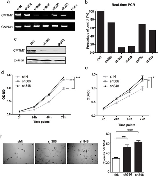
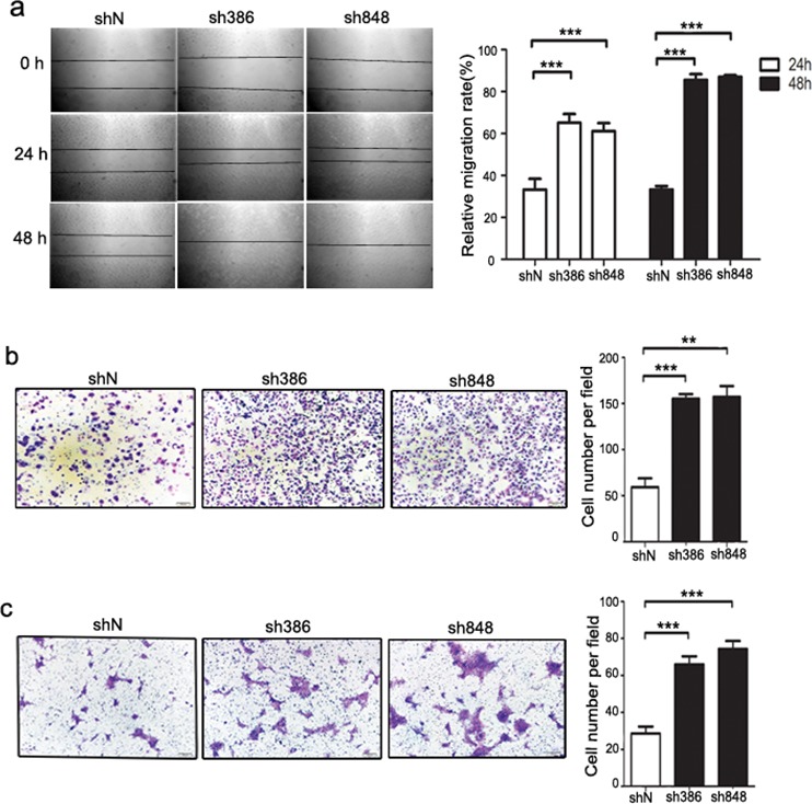
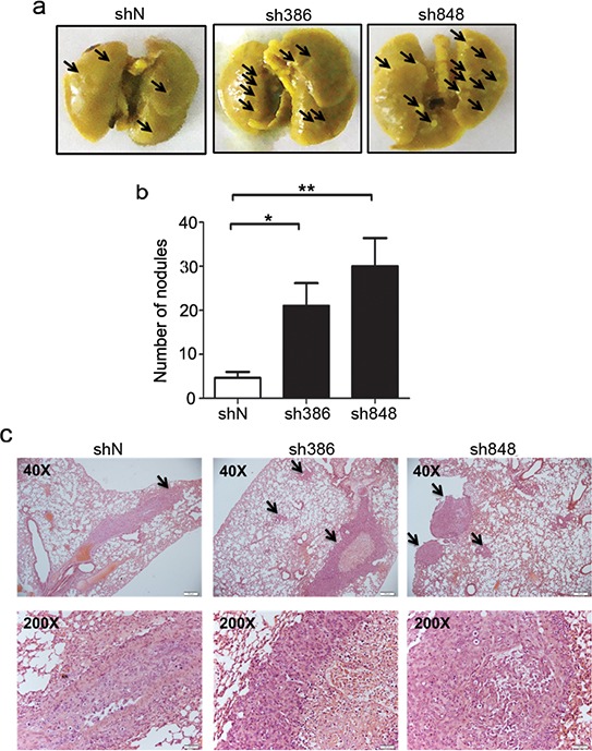
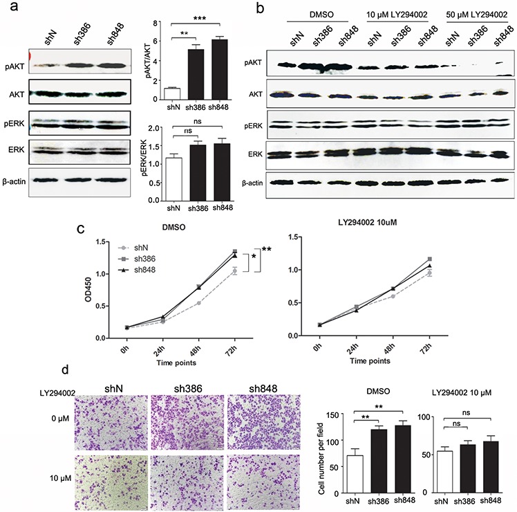
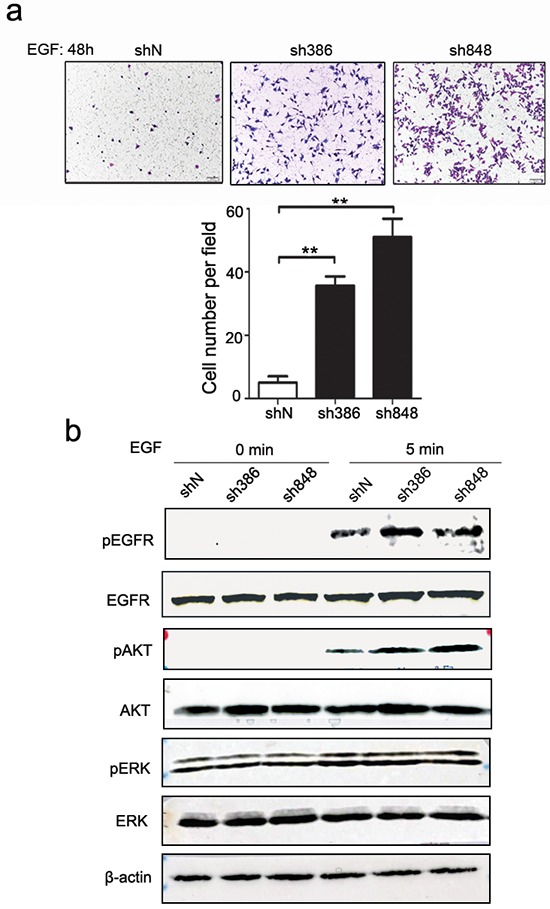
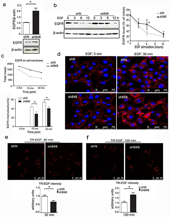
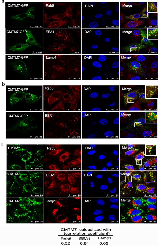
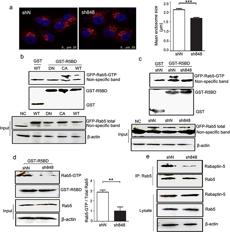
The dysregulation of epidermal growth factor receptor (EGFR) signaling has been well documented to contribute to the progression of non-small cell lung cancer (NSCLC), the leading cause of cancer death in the world. EGF-stimulated EGFR activation induces receptor internalization and degradation, which plays an important role in EGFR signaling. This process is frequently deregulated in cancer cells, leading to enhanced EGFR levels and signaling. Our previous study on CMTM7 is only limited to a brief description of the relationship of overexpressed CMTM7 with EGFR-AKT signaling. The biological functions of endogenous CMTM7 and its molecular mechanism remained unclear. In this study, we show that the stable knockdown of CMTM7 augments the malignant potential of NSCLC cells and enhances EGFR-AKT signaling by decreasing EGFR internalization and degradation. Mechanistically, CMTM7 knockdown reduces the activation of Rab5, a protein known to be required for early endosome fusion. In NSCLC, the loss of CMTM7 would therefore serve to sustain aberrant EGFR-mediated oncogenic signaling. Together, our findings highlight the role of CMTM7 in the regulation of EGFR signaling in tumor cells, revealing CMTM7 as a novel molecule related to Rab5 activation.

Keywords: AKT; CMTM7; EGFR; NSCLC; Rab5.


Conflict of interest statement

CONFLICTS OF INTEREST

The authors declare that there are no conflicts of interest.

Figures

Figure 1
Figure 1. CMTM7 knockdown promotes A549 and HCC827 cell proliferation
a, b. A549 cells were transfected with five distinct lentiviral CMTM7 shRNAs, named according to the last three numbers of the cloning item—sh208, 386, 848, 930 and 938—or the matching control non-targeting shRNA (shN). CMTM7 mRNA expression was analyzed using semi-quantitative (a) and real-time RT-PCR (b). c. A549 cells were transfected with CMTM7 shRNA (sh386, sh848) or the matching control non-targeting shRNA (shN). CMTM7 protein expression was analyzed by western blotting. d, e. CCK8 assay of control and CMTM7-knockdown NSCLC cells, A549 (d) and HCC827 cells (e). Cell numbers were determined every 24 h. Data are expressed as the mean ± s.d (*P < 0.05, ***P < 0.001). f. Representative images of cell colonies in control and CMTM7-knockdown A549 cells (magnification, × 100). Colonies with > 100 cells were quantified. Data are presented as the mean ± s.d (**P < 0.01, ***P < 0.001).
Figure 2
Figure 2. CMTM7 knockdown enhances the migration of A549 and HCC827 cells
a. Wound-healing assay of control and CMTM7-knockdown A549 cells. Confluent monolayers of control or CMTM7-knockdown cells were wounded and incubated for the indicated times. The relative migration rate at each time point (t) was calculated of changed mean gap distance (MGD) relative to the initial MGD (T0) with the formula [MGD(T0)- MGD(t)/MGD(T0)]x100%. Data are representative of three independent experiments (magnification, × 100) and are presented as the mean ± s.d (***P < 0.001). b, c. Transwell migration assay of control and CMTM7-knockdown NSCLC cells, A549 (b) and HCC827 cells (c). Photos were taken after 24 h of incubation, and the cells were stained with crystal violet (magnification, × 100). The graph indicates the mean ± s.d and P values of the number of cells per five random high-power fields (magnification, × 400) counted from three independent experiments (**P < 0.01, ***P < 0.001).
Figure 3
Figure 3. CMTM7 knockdown enhances lung metastasis of A549 cells
a. Representative images of mouse lungs. Arrows indicate pulmonary metastatic tumor nodules. b. Quantitative results (6 mice per group) of pulmonary metastatic tumor nodules at 6 weeks after injection. Data are expressed as the mean ± s.d (**P < 0.01). c. Representative hematoxylin and eosin-stained lung sections containing metastatic foci. Pictures were taken under a microscope at × 40. Arrows indicate pulmonary metastatic tumor.
Figure 4
Figure 4. Effects of CMTM7 knockdown on growth and migration via PI3K/AKT-dependent signaling
a. CMTM7 knockdown enhances AKT phosphorylation. Protein lysates of control and CMTM7-knockdown cells were immunoblotted with antibodies as indicated. The normalized densitometry data using ImageJ software are the means of three independent experiments and are presented as the mean ± s.d (**P < 0.01, ***P < 0.001; ns, not significant). b. The inhibiting efficiency of LY294002 on PI3K/Akt signaling was tested by western blotting. c. Control and CMTM7-knockdown A549 cells were treated with LY294004 (10 μM) or DMSO as a vehicle control, and cell proliferation was analyzed via CCK8 assay. Data are representative of three independent experiments. Data are expressed as the mean ± s.d (*P < 0.05, **P < 0.01). d. Control and CMTM7-knockdown A549 cells were treated with LY294004 (10 μM) or DMSO, and cell migration was analyzed via Transwell migration assay. Photos were taken after 24 h of incubation, and the cells were stained with crystal violet (magnification, × 100). The graph indicates the mean ± s.d and P values of the number of cells per five random high-power fields (magnification, × 400) counted from three independent experiments (**P < 0.01; ns, not significant).
Figure 5
Figure 5. CMTM7 knockdown results in enhanced EGF-induced migration and signaling
a. Effects of CMTM7 knockdown on EGF-induced migration via Transwell assay. Photos were taken after 48 h, and cells were stained with crystal violet (magnification, × 100). The graph indicates the mean ± s.d and P values of the number of cells per five random high-power fields (magnification, × 400) counted from three independent experiments (**P < 0.01, ***P < 0.001). b. The effect of CMTM7 knockdown on EGFR-dependent signaling was determined using western blotting of EGF-exposed cells for phospho-EGFR, phospho-AKT, and phospho-ERK. β-actin was used as an internal control.
Figure 6
Figure 6. CMTM7 knockdown attenuates EGFR internalization and delays its degradation
a. Control and CMTM7-knockdown A549 cells were cultured in complete medium, and western blots of the cell lysates were probed with antibodies against the indicated proteins. The normalized densitometry data using ImageJ software are the means of three independent experiments and are presented as the mean ± s.d (*P < 0.05). b. Control and CMTM7-knockdown A549 cells were pretreated with cycloheximide (CHX) (100 μg/ml) for 1 h prior to treatment with EGF (100 ng/ml) in the presence of CHX for the indicated times, and the cell lysates were subjected to immunoblotting with the indicated antibodies. The normalized densitometry data using ImageJ software are means of three independent experiments and are presented as the mean ± s.d (*P < 0.05). c. FACS analysis of EGFR surface levels following time-dependent EGF stimulation of CMTM7-knockdown A549 cells. EGFR internalization was quantified via mean fluorescence intensity. The graph shows the percentage of internalization at indicated time points. d. EGFR immunofluorescence analysis of CMTM7-knockdown A549 cells following EGF stimulation. Control and CMTM7-knockdown A549 cells were plated on coverslips, starved for 16 h, stimulated with 100 ng/ml EGF for 30 min, fixed, blocked and incubated with a rabbit anti-EGFR polyclonal antibody and TRITC-conjugated secondary antibody. The cells were washed prior to analysis via confocal microscopy. e, f. Control and CMTM7-knockdown A549 cells were treated with 1000 ng/ml Texas Red-EGF for 30 min at 4°C, followed by additional incubation for 30 (e) or 120 min (f) at 37°C. In each case, the fluorescence intensity of Texas Red-EGF was measured in 50 cells. Texas Red-EGF fluorescence intensity was quantified using LEICA QWin software and is presented as the mean ± s.d (*P < 0.05).
Figure 7
Figure 7. CMTM7 localizes to Rab5 and EEA1-positive microdomains on the limiting membrane of early endosomes
a. A549 cells were transfected with CMTM7-GFP for 24 h prior to fixation in 3% PFA and immunostained with antibodies against Rab5, EEA1 or Lamp1. Insets show magnification of colocalization of CMTM7-GFP with early endosomes. b. Insets show that CMTM7-GFP localizes to Rab5 and EEA1-positive “doughnut-shaped” compartments characteristic of unusually large endosomes. c. Endogenous CMTM7 localizes to early endosome and enlarged early endosome. Insets show magnification of CMTM7 induced enlarged endosomes. All endocytic markers are shown in red. Nuclei are visualized by DAPI (blue). The yellow color indicates colocalization. Bar, 25 μm. The Pearson's and Manders' overlap coefficients were derived with LEICA QWin software and are the average of ten individual cells.
Figure 8
Figure 8. CMTM7 knockdown inhibits endosome fusion by reducing Rab5 activation
a. Control and CMTM7-knockdown A549 cells were fixed in 3% PFA and stained with an anti-EEA1 (red) antibody. The different sizes of EEA1-positive endosomes were captured by confocal microscopy. Nuclei are shown in blue. Bar, 25 μm. LEICA QWin software was used to automatically measure the size of each EEA1-positive spot (***P < 0.001). b. HEK293T cells were transfected with GFP-tagged Rab5-WT, Rab5-DN, Rab5-CA, or nothing (negative control, NC). At 36 h post-transfection, the cell lysates were subjected to pull-down with GST or GST-R5BD beads. Western blots of precipitates and total cell lysates were probed with GFP antibody. c. Control and CMTM7-knockdown A549 cells were transfected with GFP-tagged Rab5-WT. After 36 h, the cells were subjected to a similar procedure and analysis. d. The lysates of control and CMTM7-knockdown A549 cells were subjected to GST or GST-R5BD pull-down, and endogenous Rab5-GTP levels were analyzed via western blotting. Relative Rab5-GTP levels are shown on the right. Data are presented as the means of three independent experiments (**P < 0.01). e. Control and CMTM7-knockdown A549 cells were lysed, and Rab5 was immunoprecipitated. The immunoprecipitated proteins were resolved on SDS-PAGE, and the levels of Rab5 and Rabaptin5 were detected by western blotting. Data are representative of three independent experiments.

References

    1. Han W, Lou Y, Tang J, Zhang Y, Chen Y, Li Y, Gu W, Huang J, Gui L, Tang Y, Li F, Song Q, Di C, Wang L, Shi Q, Sun R, et al. Molecular cloning and characterization of chemokine-like factor 1 (CKLF1), a novel human cytokine with unique structure and potential chemotactic activity. The Biochemical journal. 2001;357:127–135. - PMC - PubMed
    1. Han W, Ding P, Xu M, Wang L, Rui M, Shi S, Liu Y, Zheng Y, Chen Y, Yang T, Ma D. Identification of eight genes encoding chemokine-like factor superfamily members 1–8 (CKLFSF1-8) by in silico cloning and experimental validation. Genomics. 2003;81:609–617. - PubMed
    1. Li T, Zhong J, Chen Y, Qiu X, Zhang T, Ma D, Han W. Expression of chemokine-like factor 1 is upregulated during T lymphocyte activation. Life sciences. 2006;79:519–524. - PubMed
    1. Zhang Y, Wang JY, Han W. A role for CMTM7 in BCR expression and survival in B-1a but not B-2 cells. International immunology. 2014;26:47–57. - PubMed
    1. Miyazaki A, Yogosawa S, Murakami A, Kitamura D. Identification of CMTM7 as a transmembrane linker of BLNK and the B-cell receptor. PloS one. 2012;7:e31829. - PMC - PubMed

Publication types

MeSH terms