. 2015 Nov 3;4(11):e263.
doi: 10.1038/mtna.2015.36.

Preclinical Development of a Subcutaneous ALAS1 RNAi Therapeutic for Treatment of Hepatic Porphyrias Using Circulating RNA Quantification

Affiliations

Preclinical Development of a Subcutaneous ALAS1 RNAi Therapeutic for Treatment of Hepatic Porphyrias Using Circulating RNA Quantification

Amy Chan et al. Mol Ther Nucleic Acids. .

Abstract

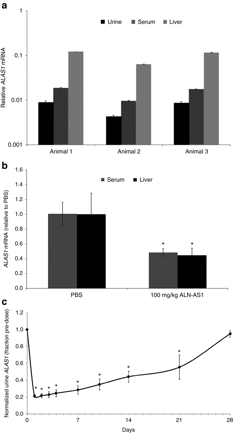
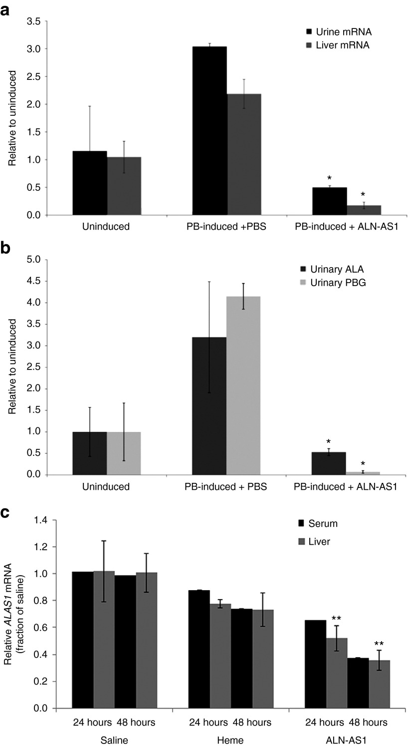
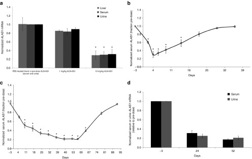
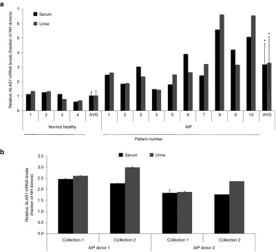
The acute hepatic porphyrias are caused by inherited enzymatic deficiencies in the heme biosynthesis pathway. Induction of the first enzyme 5-aminolevulinic acid synthase 1 (ALAS1) by triggers such as fasting or drug exposure can lead to accumulation of neurotoxic heme intermediates that cause disease symptoms. We have demonstrated that hepatic ALAS1 silencing using siRNA in a lipid nanoparticle effectively prevents and treats induced attacks in a mouse model of acute intermittent porphyria. Herein, we report the development of ALN-AS1, an investigational GalNAc-conjugated RNAi therapeutic targeting ALAS1. One challenge in advancing ALN-AS1 to patients is the inability to detect liver ALAS1 mRNA in the absence of liver biopsies. We here describe a less invasive circulating extracellular RNA detection assay to monitor RNAi drug activity in serum and urine. A striking correlation in ALAS1 mRNA was observed across liver, serum, and urine in both rodents and nonhuman primates (NHPs) following treatment with ALN-AS1. Moreover, in donor-matched human urine and serum, we demonstrate a notable correspondence in ALAS1 levels, minimal interday assay variability, low interpatient variability from serial sample collections, and the ability to distinguish between healthy volunteers and porphyria patients with induced ALAS1 levels. The collective data highlight the potential utility of this assay in the clinical development of ALN-AS1, and in broadening our understanding of acute hepatic porphyrias disease pathophysiology.


Figures

Figure 1
Figure 1
Detection of ALAS1 RNA from rodent serum or urine and correlation with RNAi-mediated gene silencing in liver. (a) Relative levels of ALAS1, as measured by quantitative polymerase chain reaction in RNA isolated from filtered, centrifuged rat serum. Values are relative to GAPDH levels; error bars, SD in experimental replicates. (b) Silencing of ALAS1 mRNA in rat liver and serum 72 hours after subcutaneous administration of 10 mg/kg ALN-AS1 or phosphate-buffered saline (PBS) control (vehicle). Levels of ALAS1 were normalized to GAPDH levels. ALAS1 liver and serum mRNA levels from individual animals were normalized to those from a group average of three PBS-treated livers and serum, respectively. Each graphical data point represents the percent remaining ALAS1 mRNA relative to the group average PBS-treated ALAS1 mRNA ± SD of the group. *Significance was determined by Student's t-test (P < 0.05). (c) Time course of ALAS1 mRNA silencing normalized to GAPDH monitored in urine following subcutaneous administration of 10 mg/kg ALN-AS1. ALAS1 transcript from individual animals was normalized to their respective individual predose ALAS1 level at each timepoint. Each graphical data point represents the fraction remaining ALAS1 relative to predose for the group average of four animal samples assayed in technical triplicates ± the standard deviation of the group. *Significance was determined by Student's t-test (P < 0.001).
Figure 2
Figure 2
Silencing of liver ALAS1 mRNA upon prophylactic and acute administration of ALN-AS1 quantified by circulating mRNA levels in rodent models of acute intermittent porphyria. Wild-type rats were administered ALN-AS1 or phosphate-buffered saline (PBS) prophylactically, twice weekly for 3 weeks. Six days prior to the final dose, all animals were i.v. administered Porphobilinogen deaminase siRNA. This was followed by a challenge with 4-daily phenobarbital (PB) injections to induce ALAS1 levels in all animals, excluding the “Uninduced” cohort. Liver and urine samples were collected 24 hours after the final PB injection. (a) ALAS1 mRNA levels in liver and urine and (b) urinary ALA and porphobilinogen concentrations were measured. Data are presented as mean ± SD (n = 3). *Significance was determined by Student's t-test (P < 0.05). (c) Acute intermittent porphyria mice were biochemically induced with four daily doses of 3,5-diethoxycarbonyl-1,4-dihydro-2,4,6-trimethylpyridine and PB and treated with a single subcutaneous injection of ALN-AS1 (20 mg/kg), saline, or intravenous heme (4.0 mg/kg) (n = 7–10 per group) ~4 hours after the third PB dose. The heme treatment group was administered a second dose of heme 24 hours after the first. Terminal blood collections were made from cohorts sacrificed at 24 and 48 hours post-treatment (n = 7–10/time point), at which times livers were harvested from a subset of mice (n = 4). Serum and liver transcript levels were determined and are presented as mean ± SD. **Significance for ALN-AS1-treated liver samples relative to Heme-treated animals was determined by Student's t-test (P < 0.05).
Figure 3
Figure 3
Evaluation of ALAS1 mRNA silencing in circulation and liver in nonhuman primates treated with ALN-AS1. (a) Silencing of liver, serum, and urine ALAS1 mRNA normalized to GAPDH in cynomolgus monkey (M. fascicularis) 96 hours after subcutaneous administration of indicated doses of ALN-AS1. ALAS1 liver mRNA levels from individual animals were normalized to those from a group average of three PBS-treated liver biopsies. Each graphical data point represents the percent remaining ALAS1 mRNA relative to the group average PBS-treated ALAS1 mRNA ± SD of the group. ALAS1 serum and urine transcript from individual animals was normalized to their respective individual predose ALAS1 level. Each graphical data point represents the fraction remaining ALAS1 relative to predose for the group average of three animal samples assayed in technical triplicates ± the standard deviation of the group. *Significance relative to PBS-treated samples for liver transcript and relative to pre-ALN-AS1 treatment for serum and urine was determined by Student's t-test (P < 0.05). (b) Time course of ALAS1 mRNA silencing in cynomolgus monkey serum following a single subcutaneous administration of ALN-AS1 at 10 mg/kg. ALAS1 serum transcript from individual animals was normalized to their respective individual predose ALAS1 level at each timepoint. Each graphical data point represents the fraction remaining ALAS1 relative to predose for the group average of three animal samples assayed in technical triplicates ± the standard deviation of the group. (c) Time course of ALAS1 silencing in cynomolgus monkey serum following subcutaneous administration of 5 mg/kg once weekly for 8 weeks of ALN-AS1 and (d) comparison between serum and urine ALAS1 levels following subcutaneous administration of 5 mg/kg once weekly for 8 weeks of ALN-AS1. Levels of ALAS1 were normalized to those of GAPDH. ALAS1 serum and urine transcript from individual animals was normalized to their respective individual predose ALAS1 level at each timepoint. Each graphical data point represents the fraction remaining ALAS1 relative to pre-dose for the group average of three animal samples assayed in technical triplicates ± the standard deviation of the group. *Significance relative to pre-ALN-AS1 treatment for serum and urine was determined by Student's t-test (P < 0.05).
Figure 4
Figure 4
Assessment of ALAS1 transcript levels in normal and acute intermittent porphyria (AIP) human donor-matched serum and urine samples. (a) ALAS1 mRNA levels in filtered AIP donor-matched serum and urine (n = 10) relative to those from normal healthy volunteers (n = 4). Values are relative to GAPDH levels; error bars, SD in experimental replicates. *Significance was determined by Student's t-test (P < 0.05). (b) Comparison of ALAS1 mRNA levels in AIP donor-matched, circulating serum and urine collected at two different time points, 3 weeks apart (n = 2). Values are relative to GAPDH levels; error bars, SD in experimental replicates.

References

    1. Anderson, KE, Sassa, S, Bishop, DF, Desnick, RJ (2001). Disorders of heme biosynthesis: X linked sideroblastic anemia and the porphyrias. In: Scriver CR, Beaudet AL, Sly WS, Valle D (eds.). The Metabolic and Molecular Bases of Inherited Disease. McGraw-Hill, New York. pp. 2961–3062.
    1. Granick, S (1963). Induction of the synthesis of delta-aminolevulinic acid synthetase in liver parenchyma cells in culture by chemical that induce acute porphyria. J Biol Chem 238: 2247–2249. - PubMed
    1. de Matteis, F (1967). Disturbances of liver porphyrin metabolism caused by drugs. Pharmacol Rev 19: 523–557. - PubMed
    1. Sassa, S, Bradlow, HL and Kappas, A (1979). Steroid induction of delta-aminolevulinic acid synthase and porphyrins in liver. Structure-activity studies and the permissive effects of hormones on the induction process. J Biol Chem 254: 10011–10020. - PubMed
    1. Miller, LK and Kappas, A (1974). The effect of progesterone on activities of delta-aminolevulinic acid synthetase and delta-aminolevulinic acid dehydratase in estrogen-primed avian oviduct. Gen Comp Endocrinol 22: 238–244. - PubMed