Review
. 2015 Oct 29;16(11):25959-81.
doi: 10.3390/ijms161125939.

Energy Metabolism of the Brain, Including the Cooperation between Astrocytes and Neurons, Especially in the Context of Glycogen Metabolism

Affiliations
Review

Energy Metabolism of the Brain, Including the Cooperation between Astrocytes and Neurons, Especially in the Context of Glycogen Metabolism

Anna Falkowska et al. Int J Mol Sci. .

Abstract

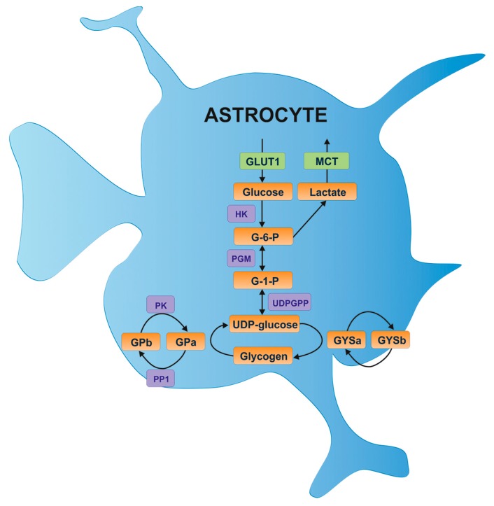
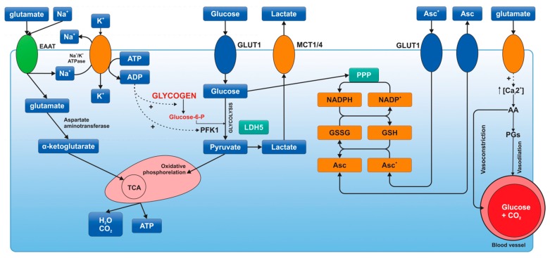
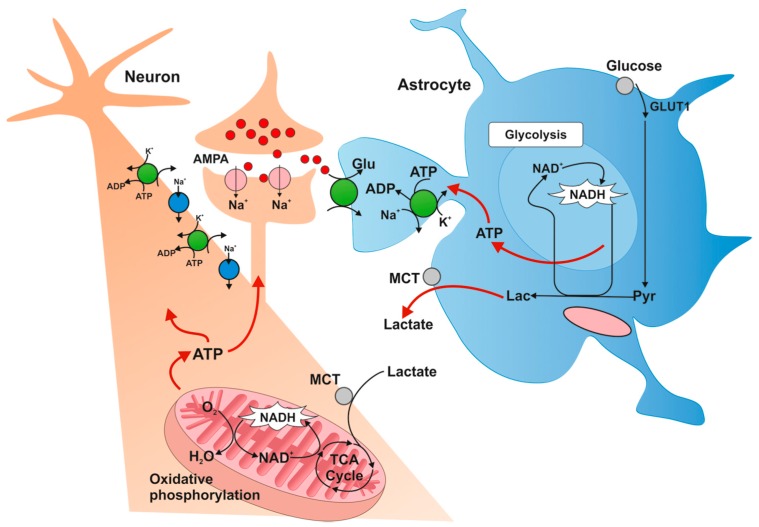
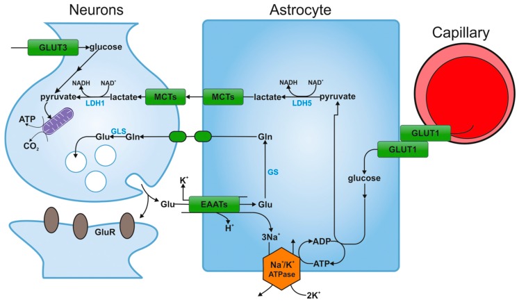
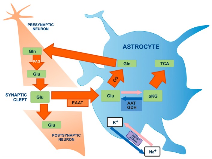
Glycogen metabolism has important implications for the functioning of the brain, especially the cooperation between astrocytes and neurons. According to various research data, in a glycogen deficiency (for example during hypoglycemia) glycogen supplies are used to generate lactate, which is then transported to neighboring neurons. Likewise, during periods of intense activity of the nervous system, when the energy demand exceeds supply, astrocyte glycogen is immediately converted to lactate, some of which is transported to the neurons. Thus, glycogen from astrocytes functions as a kind of protection against hypoglycemia, ensuring preservation of neuronal function. The neuroprotective effect of lactate during hypoglycemia or cerebral ischemia has been reported in literature. This review goes on to emphasize that while neurons and astrocytes differ in metabolic profile, they interact to form a common metabolic cooperation.

Keywords: astrocytes; brain; brain energy metabolism; glucose; glycogen; neurons.


Figures

Figure 1
Figure 1
Glucose entry and glycogen formation in astrocytes [13]. Glucose is transported via the glucose transporter 1 (GLUT1) and possibly the insulin-sensitive glucose transporter 4 (GLUT4). Glucose is phosphorylated by hexokinase (HK) to glucose-6-phosphate (G-6-P), which is subsequently converted to glucose-1-phosphate (G-1-P) by phosphoglucomutase (PGM) and then to UDP glucose by UDP glucose pyrophosphate (UDPGPP). The UDP glucose continues on to glycogen synthesis via the actions of glycogen synthase (GYS), which can exist in two forms: the active dephosphorylated form (GYSa) or the inactive phosphorylated form (GYSb). Protein phosphatase 1 (PP1) converts GYSb to active GYSa via the regulatory subunit Protein Targeting to Glycogen (PTG), resulting in glycogen formation. Glycogen is broken down by glycogen phosphorylase (GP), which similar to glycogen synthase exists in two forms: the active phosphorylated form (GPa), or the inactive dephosphorylated form (GPb). Phosphorylase kinase (PK) dephosphorylates GPb to the active form. A major glycogen-derived product is lactate, which is transported into the extracellular space via monocarboxylate transporters (MCT).
Figure 2
Figure 2
Metabolic activation of astrocytes [40]. Synaptic activity produces an increase in extracellular K+, which stimulates Na+K+-ATPase by binding of its extracellular K+-sensitive site. The excitatory neurotransmitter glutamate is taken up by astrocytes through excitatory amino acid transporters. This kind of transport produces an increase in intracellular Na+, which stimulates Na+K+-ATPase by binding of its intracellular Na+-sensitive site. Na+K+-ATPase activation produces a decrease in ATP/ADP ratio, thus glycolysis and glycogenolysis activation. In addition, glucose is oxidized by pentose phosphate pathway (PPP) to produce NADPH and to maintain the redox balance, reducing glutathione and ascorbic acid (Asc). Ascorbic acid released by astrocytes is taken up by neurons to protect themselves from oxidant species (ascorbic acid is oxidized in neurons). Oxidized ascorbic acid (dehydroascorbic acid, Asc+) is released from neurons and taken up by astrocytes through GLUT1. In astrocytes, glutamate is able to bind ionotropic receptors, which are predominantly calcium channels. This Ca2+ increase cooperates with Krebs cycle (TCA) activation and produces arachidonic acid (AA) and prostaglandin (PGs), which stimulate the constriction and dilation of capillaries, respectively. ADP: adenosine diphosphate; ATP: adenosine triphosphate; EAAT: excitatory amino-acid transporter; GLUT: glucose transporter; G-6-P: glucose 6-phosphate; GSSG: glutathione disulfide; GSH: glutathione; LDH: lactate dehydrogenase; MCT: monocarboxylate transporter; Na+K+-ATPase: sodium-potassium pump; NADP+: nicotinamide adenine dinucleotide phosphate (NADPH-reduced form of NADP); PFK1: phosphofructokinase-1; TCA: Krebs cycle.
Figure 3
Figure 3
Metabolic profile of astrocytes [16]. Glucose is transported to astrocytes through GLUT1 transporter and then metabolized to lactate. Lactate is transported outside astrocytes and taken up by neurons by monocarboxylate transporters (MCTs). Intracellular lactate in neurons is oxidized to pyruvate and metabolized along the oxygen pathway. GLUT1: glucose transporter-1; MCT: monocarboxylate transporter; TCA: Krebs cycle; Pyr: pyruvate; Lac: lactate.
Figure 4
Figure 4
The hypothesis of astrocyte-neuron lactate shuttle [16]. Glucose is transported from blood vessels to astrocytes by endothelial cells. After entering the astrocyte via GLUT1, part of the glucose is metabolized to lactate via pyruvate by the isoenzyme of lactate dehydrogenase (LDH5). Then lactate is transported outside the astrocyte through the MCT transporter and is captured by neurons, also via the MCT transporter. Intracellular lactate in the neuron is oxidized to pyruvate by the other isoenzyme of lactate hydrogenase (LDH1) and is metabolized along the oxygen pathway. Glucose may be also transported directly into neuronal cells and penetrates into these cells via GLUT3. GLUT1, GLUT3: glucose transporters; MCT: monocarboxylate transporter; TCA: Krebs cycle; GS: glutamine synthetase; Glu: glutamate; Gln: glutamine; GluR: receptor for glutamate; EAATs: excitatory amino-acid transporters; GLS: glutaminase; LDH1, LDH5: isoenzymes of lactate dehydrogenase.
Figure 5
Figure 5
Glutamate-glutamine cycle [77]. Glutamate (Glu) molecules are released from the pre-synaptic neuron into the synaptic cleft where they can stimulate glutamate receptors on the post-synaptic neuron. Glutamate also penetrates the synaptic gap towards astrocytes. Glu is mainly transported to astrocytes by Na+-dependent excitatory amino-acid activating transporters (EAATs). This leads to the occurrence of astrocyte ion gradients of Na+, which stimulates Na+/K+ ATPase in order to restore proper ionic concentrations. Glutamate is transformed into: glutamine (Gln) by means of glutamine synthase enzyme (GS) or to α-ketoglutarate (α-KG) by means of glutamate dehydrogenase (GDH) or aspartate aminotransferase (AAT), in order to conduct further oxygen metabolism in the TCA cycle (Krebs cycle). Glutamine is transferred to neurons in order to conduct further glutamate production with the participation of phosphate-activated glutaminase (PAG).

References

    1. Secher N.H., Quistorff B. Brain glucose and lactate uptake during exhaustive exercise. J. Physiol. 2005;568:3. doi: 10.1113/jphysiol.2005.095786. - DOI - PMC - PubMed
    1. Cataldo A.M., Broadwell R.D. Cytochemical identification of cerebral glycogen and glucose-6-phosphatase activity under normal and experimental conditions. II. Choroid plexus and ependymal epithelia, endothelia and pericytes. J. Neurocytol. 1986;15:511–524. doi: 10.1007/BF01611733. - DOI - PubMed
    1. Nehlig A., Pereira de Vasconcelos A. Glucose and ketone body utilization by the brain of neonatal rats. Prog. Neurobiol. 1993;40:163–221. doi: 10.1016/0301-0082(93)90022-K. - DOI - PubMed
    1. Huang Y.H., Bergles D.E. Glutamate transporters bring competition to the synapse. Curr. Opin. Neurobiol. 2004;14:346–352. doi: 10.1016/j.conb.2004.05.007. - DOI - PubMed
    1. Matthias K., Kirchhoff F., Seifert G., Huttmann K., Matyash M., Kettenmann H., Steinhauser C. Segregated expression of AMPA-type glutamate receptors and glutamate transporters defines distinct astrocyte populations in the mouse hippocampus. J. Neurosci. 2003;23:1750–1758. - PMC - PubMed

Publication types