. 2015 Nov 3;10(11):e0141862.
doi: 10.1371/journal.pone.0141862. eCollection 2015.

Adipose Mesenchymal Stem Cell Secretome Modulated in Hypoxia for Remodeling of Radiation-Induced Salivary Gland Damage

Affiliations

Adipose Mesenchymal Stem Cell Secretome Modulated in Hypoxia for Remodeling of Radiation-Induced Salivary Gland Damage

Hye-Young An et al. PLoS One. .

Abstract

Background and purpose: This study was conducted to determine whether a secretome from mesenchymal stem cells (MSC) modulated by hypoxic conditions to contain therapeutic factors contributes to salivary gland (SG) tissue remodeling and has the potential to improve irradiation (IR)-induced salivary hypofunction in a mouse model.

Materials and methods: Human adipose mesenchymal stem cells (hAdMSC) were isolated, expanded, and exposed to hypoxic conditions (O2 < 5%). The hypoxia-conditioned medium was then filtered to a high molecular weight fraction and prepared as a hAdMSC secretome. The hAdMSC secretome was subsequently infused into the tail vein of C3H mice immediately after local IR once a day for seven consecutive days. The control group received equal volume (500 μL) of vehicle (PBS) only. SG function and structural tissue remodeling by the hAdMSC secretome were investigated. Human parotid epithelial cells (HPEC) were obtained, expanded in vitro, and then irradiated and treated with either the hypoxia-conditioned medium or a normoxic control medium. Cell proliferation and IR-induced cell death were examined to determine the mechanism by which the hAdMSC secretome exerted its effects.

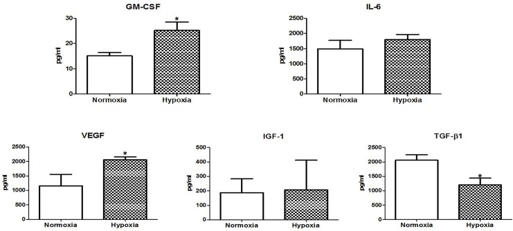
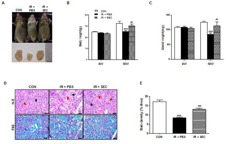
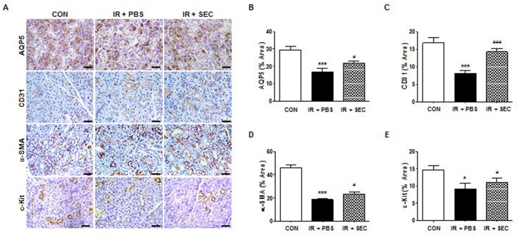
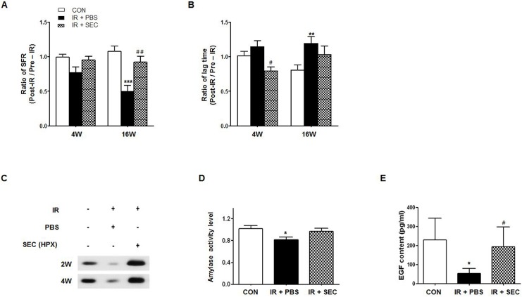
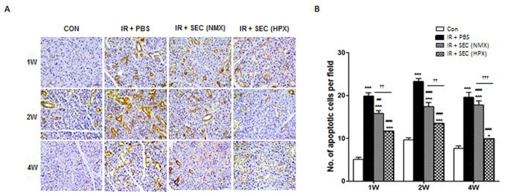
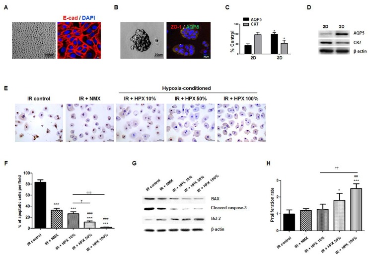
Results: The conditioned hAdMSC secretome contained high levels of GM-CSF, VEGF, IL-6, and IGF-1. Repeated systemic infusion with the hAdMSC secretome resulted in improved salivation capacity and increased levels of salivary proteins, including amylase and EGF, relative to the PBS group. The microscopic structural integrity of SG was maintained and salivary epithelial (AQP-5), endothelial (CD31), myoepithelial (α-SMA) and SG progenitor cells (c-Kit) were successfully protected from radiation damage and remodeled. The hAdMSC secretome strongly induced proliferation of HPEC and led to a significant decrease in cell death in vivo and in vitro. Moreover, the anti-apoptotic effects of the hAdMSC secretome were found to be promoted after hypoxia-preconditioning relative to normoxia-cultured hAdMSC secretome.

Conclusion: These results show that the hAdMSC secretome from hypoxic-conditioned medium may provide radioprotection and tissue remodeling via release of paracrine mediators.


Conflict of interest statement

Competing Interests: K-Stem Co., Ltd. provided support in the form of experimental cell sources. There are no patents, products in development, or marketed products to declare. This does not alter the authors' adherence to all the PLOS ONE policies on sharing data and materials.

Figures

Fig 1
Fig 1. Secretome analysis.
Concentrations of cytokine and growth factors released by hAdMSC were measured after hypoxia preconditioning with O2 at < 5% for 6 hours using multiplex antibody arrays. Data displayed are from the hAdMSC secretomes generated under normoxia and hypoxia. Mann-Whitney test (A) GM-CSF, P = 0.048, (B) IL-6, P = 0.3914, (C) VEGF, P = 0.044, (D) IGF-1, P = 0.936; n = 3. Each sample was measured in duplicate.
Fig 2
Fig 2. Macro- and micro-morphological evaluation.
(A) External appearance and dissected SGs of mice in each group were photographed at 16 weeks after IR. (B and C) Body weight and dissected glandular weight were measured at 4 and 16 weeks after IR. (D) Representative histological pictures of H-E staining (upper) and PAS staining (lower) from three groups at 16 weeks post-IR are presented. Red arrows indicate ductal structures and stars mark acinar structures. Scale bars represent 50 μm. (E) Densities upon PAS staining were measured using a software program to calculate pixels of purple stained mucin-containing areas. Data are presented as the mean ± SEM. Two-way ANOVA, Bonferroni post hoc test (B and C), One-way ANOVA, Tukey’s pot hoc test (E). *, compared to CON; #, compared to IR + PBS. ***P <0.001, ##P < 0.01, and ###P < 0.001. CON, normal control group (n = 6–7); IR + PBS, PBS-treated group (n = 6); IR + SEC, secretome-treated group (n = 6).
Fig 3
Fig 3. Cytoprotective effect of hAdMSC secretome treatment.
(A) Representative images of salivary epithelial (AQP5), endothelial (CD31), myoepithelial (α-SMA), and progenitor cells (c-Kit) in the SG in the three experimental groups. Scale bars represent 50 μm. (B-E) Each staining area was measured in pixels using a software program. Data are presented as the mean area (%) ± SEM. One-way ANOVA, Tukey’s pot hoc test. *, compared to CON; #, compared to IR + PBS. *P < 0.05, ***P <0.001, #P < 0.05, and ###P < 0.001. CON, normal control group; IR + PBS, PBS-treated group; IR + SEC, secretome-treated group. Three random sections from each mouse were evaluated by a blinded researcher. The total number of slides examined from each experimental group ranged from 18 to 21.
Fig 4
Fig 4. Recovery of salivary hypofunction by hAdMSC secretome infusion.
(A) Salivary flow rate (SFR) was calculated at pre-IR and 4 and 16 weeks post-IR. The changes in SFR after IR were expressed by the ratio of post-IR SFR to pre-IR SFR (Mean ± SEM). (B) Time to salivation (lag time, LT) was measured and the ratios of post-IR LT to pre-IR LT were presented. (C) Western blotting of amylase in saliva at 2 and 4 weeks post-IR. (D) The salivary amylase activity was examined by the Assay Kit and fold changes in activity level are presented. (E) EGF contents were measured at each time point and the average contents are presented. Data are presented as the mean ± SEM. Two-way ANOVA, Bonferroni post hoc test (A and B), one-way ANOVA, Tukey’s pot hoc test (D and E). *, compared to CON; #, compared to IR + PBS. *P <0.05, **P < .01, ***P <0.001, #P < 0.05, and ##P < 0.01. CON, normal control group (n = 5–7); IR + PBS, PBS-treated group (n = 5–6); IR + SEC, secretome-treated group (n = 5–6).
Fig 5
Fig 5. hAdMSC secretome exerts cytoprotection via anti-apoptotic effects on salivary parenchymal cells in vivo.
(A) Representative images of an in vivo TUNEL assay from three experimental groups at 1, 2, and 4 weeks post-IR. Scale bars represent 50 μm. (B) The number of TUNEL-positive apoptotic cells was determined by a blinded researcher. Data are presented as the mean number of apoptotic cells per field ± SEM. Two-way ANOVA, Bonferroni post hoc test. *, compared to CON; #, compared to IR + PBS; †, compared to IR + NMX. *P <0.01, ***P <0.001, ##P < 0.01, ###P < 0.001, ††P<0.01, †††P<0.001. CON, normal control group (n = 21 sections); IR+ PBS, PBS-treated group (n = 21 sections); IR + SEC (NMX), IR + normoxic secretome-treated group (n = 15 sections); IR + SEC (HPX), hypoxia-conditioned secretome-treated group (n = 18 sections).
Fig 6
Fig 6. In vitro effect of the hAdMSC secretome on cell death and proliferation of human parotid epithelial cells (HPEC) treated with the hAdMSC secretome at concentrations of 10, 50 and 100% or a normoxic control medium.
(A–B) Light microscopic and immunofluorescent staining of HPEC. Scale bars represent 100 and 8μm in A and 100 and 20 μm in B. (C-D) Salivary epithelial genes of AQP5 and CK7 were confirmed by real-time PCR and western blot. *, compared to 2D, *P < 0.05. (E) Anti-apoptotic effect was confirmed by an in vitro TUNEL assay in HPEC. (F) The number of fragmented DNA of HPEC according to the concentration of hAdMSC-cultured medium was determined in the same manner. Data are presented as the mean number of apoptotic cells per field ± SEM. One-way ANOVA, Tukey’s pot hoc test. *, compared to 0%; #, compared to IR + PBS; †, compared to IR + NMX. ***P < 0.001, ###P < 0.001, †P < 0.05, †††P < 0.001. 0%, control standard medium. (G) IR-induced changes in Bax, cleaved caspase-3 and Bcl-2 in HPEC treated with the hypoxia-cultured hAdMSC medium at concentrations of 10, 50 and 100% or a normoxic control medium for 72 hours were evaluated using Western blot. (H) Cell proliferation of HPEC was assessed in triplicate using a CCK-8. Data are presented as the mean number of cells per field ± SEM. One-way ANOVA, Tukey’s pot hoc test. *, compared to 0%; #, compared to IR + PBS; †, compared to IR + NMX. *P < 0.05, ***P < 0.001, ##P < 0.01, ††P < 0.01; IR + NMX, IR + normoxia-cultured hAdMSC medium, IR + HPX, hypoxia-conditioned hAdMSC medium.

References

    1. Dirix P, Nuyts S, Van den Bogaert W (2006) Radiation-induced xerostomia in patients with head and neck cancer: a literature review. Cancer 107: 2525–2534. - PubMed
    1. Jensen SB, Pedersen AM, Vissink A, Andersen E, Brown CG, Davies AN, et al. (2010) A systematic review of salivary gland hypofunction and xerostomia induced by cancer therapies: prevalence, severity and impact on quality of life. Support Care Cancer 18: 1039–1060. 10.1007/s00520-010-0827-8 - DOI - PubMed
    1. Jensen SB, Pedersen AM, Vissink A, Andersen E, Brown CG, Davies AN, et al. (2010) A systematic review of salivary gland hypofunction and xerostomia induced by cancer therapies: management strategies and economic impact. Support Care Cancer 18: 1061–1079. 10.1007/s00520-010-0837-6 - DOI - PubMed
    1. Sasportas LS, Hosford DN, Sodini MA, Waters DJ, Zambricki EA, Barral JK, et al. (2013) Cost-effectiveness landscape analysis of treatments addressing xerostomia in patients receiving head and neck radiation therapy. Oral Surg Oral Med Oral Pathol Oral Radiol 116: e37–51. 10.1016/j.oooo.2013.02.017 - DOI - PMC - PubMed
    1. Azzam EI, Jay-Gerin JP, Pain D (2012) Ionizing radiation-induced metabolic oxidative stress and prolonged cell injury. Cancer Lett 327: 48–60. 10.1016/j.canlet.2011.12.012 - DOI - PMC - PubMed

Publication types

MeSH terms

LinkOut - more resources