. 2015 Nov 4:5:16064.
doi: 10.1038/srep16064.

Mitofilin and CHCHD6 physically interact with Sam50 to sustain cristae structure

Affiliations

Mitofilin and CHCHD6 physically interact with Sam50 to sustain cristae structure

Chengli Ding et al. Sci Rep. .

Abstract

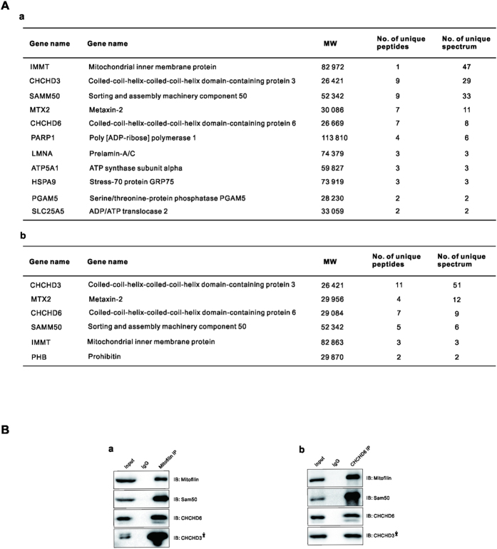
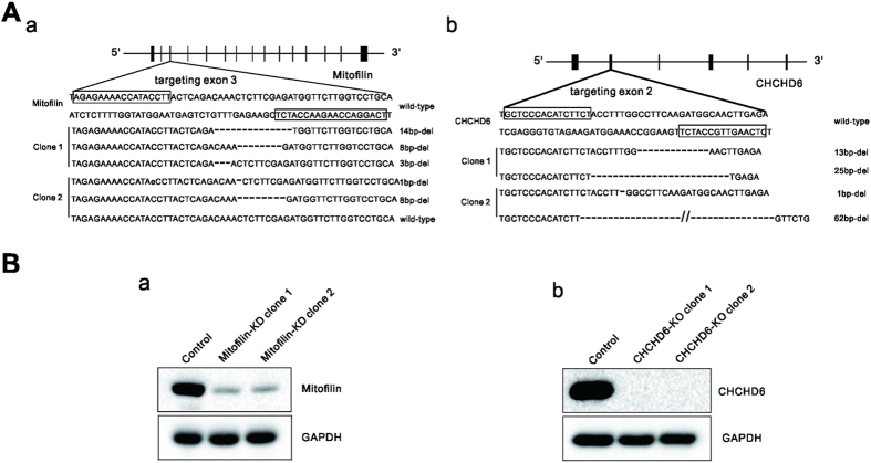
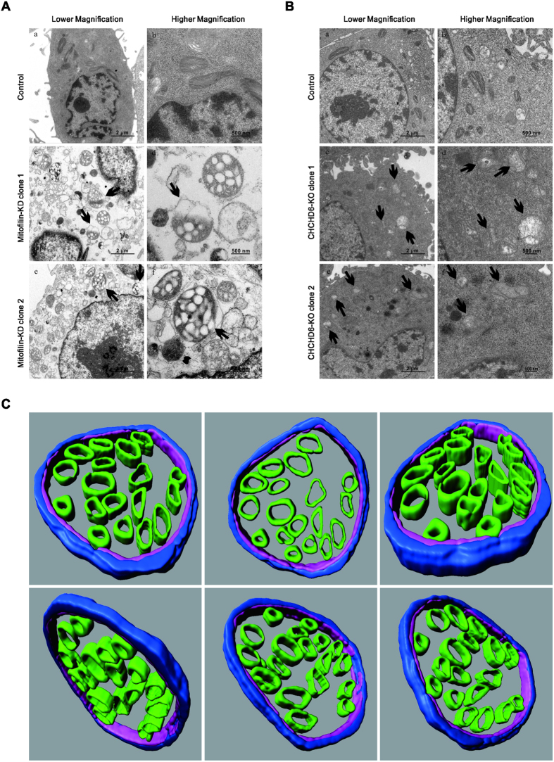
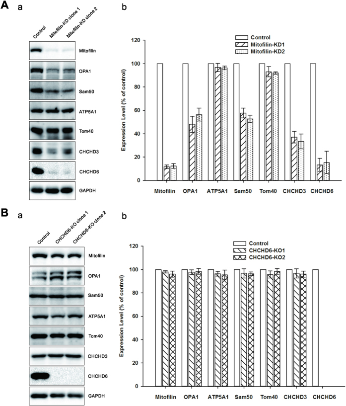
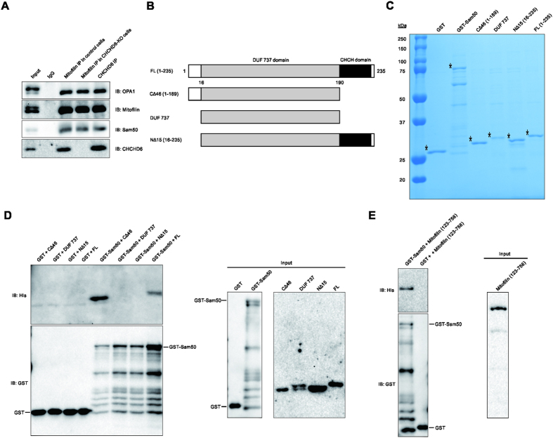
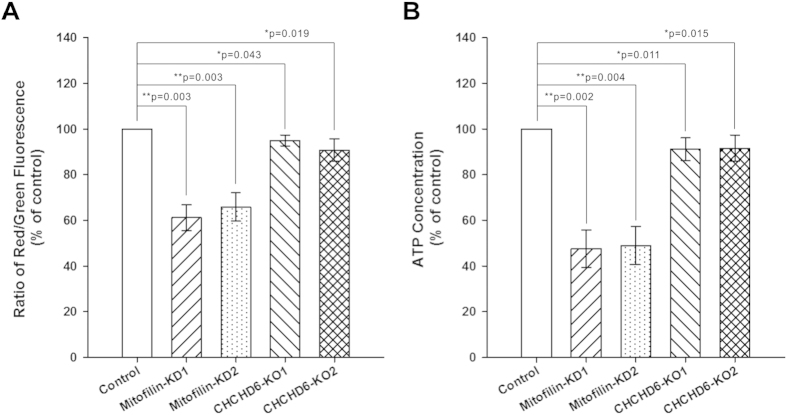
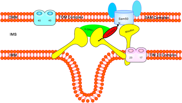
The inner mitochondrial membrane (IMM) invaginates to form cristae and the maintenance of cristae depends on the mitochondrial contact site (MICOS) complex. Mitofilin and CHCHD6, which physically interact, are two components of the MICOS. In this study, we performed immunoprecipitation experiments with Mitofilin and CHCHD6 antibodies and identified a complex containing Mitofilin, Sam50, and CHCHD 3 and 6. Using transcription activator-like effector nucleases (TALENs), we generated knockdown/knockout clones of Mitofilin and CHCHD6. Transmission electron microscopy (TEM) revealed that vesicle-like cristae morphology appeared in cell lines lacking Mitofilin, and mitochondria exhibited lower cristae density in CHCHD6-knockout cells. Immunoblot analysis showed that knockdown of Mitofilin, but not knockout of CHCHD6, affected their binding partners that control cristae morphology. We also demonstrated that Mitofilin and CHCHD6 directly interacted with Sam50. Additionally, we observed that Mitofilin-knockdown cells showed decreased mitochondrial membrane potential (ΔΨm) and intracellular ATP content, which were minimally affected in CHCHD6-knockout cells. Taken together, we conclude that the integrity of MICOS and its efficient interaction with Sam50 are indispensable for cristae organization, which is relevant to mitochondrial function.


Figures

Figure 1
Figure 1
Mitofilin, Sam50, and CHCHD 3 and 6 are in the same complex, (A) a and b show MS data for the indicated proteins from the Mitofilin and CHCHD6 IPs. The number of total peptides (Total) and total unique peptides (Unique) identified by MS are shown. (B) The IP sample of endogenous Mitofilin (a) and CHCHD6 (b) were analyzed via SDS-PAGE followed by immunoblotting (IB) with indicated antibodies. Asterisks indicated that the blot of CHCHD3 was cropped from a different gel, as CHCHD3 and CHCHD6 have similar sizes. Full-length blots/gels are presented in Supplementary Figure 3.
Figure 2
Figure 2
Generation of Mitofilin-knockdown and CHCHD6-knockout HeLa cell clones with TALENs (A) The boxes indicate the TALEN binding sites for Mitofilin, targeting exon 3 (a), and for CHCHD6, targeting exon 2 (b).Deletions in alleles of each clone are indicated. (B) Immunoblot analysis using whole-cell lysates from wild-type (WT) cells and the two Mitofilin-knockdown (a) or two CHCHD6-knockout (b) HeLa cell lines. Full-length blots/gels are presented in Supplementary Figure 4.
Figure 3
Figure 3. Altered cristae morphology caused by Mitofilin knockdown or CHCHD6 knockout.
(A,B) Electron microscopy of mitochondria in control and Mitofilin-knockdown (A) or CHCHD6-knockout cells (B). The black arrows indicate disrupted mitochondria. (C) Electron tomography of mitochondrial morphological changes in Mitofilin-knockdown cells. The OMM is depicted in light blue, the IBM is shown in pink, and cristae are shown in green. These images are rotations of surface-rendered views of tomographic reconstructions of mitochondria.
Figure 4
Figure 4. Steady-state levels several MICOS components in Mitofilin-knockdown or CHCHD6-knockout cells.
Equal amounts of protein samples in control cells and Mitofilin knockdown cells (A) or CHCHD6 knockout cells (B) were analyzed via SDS-PAGE followed by immunoblot with indicated antibodies. The values represent the average protein expression ± SD from three independent experiments. GAPDH was used as a loading control. Full-length blots/gels are presented in Supplementary Figure 5.
Figure 5
Figure 5. CHCHD6 interacts with OPA1 and directly interacts with Sam50.
(A) The IP samples of Mitofilin and CHCHD6 were analyzed via SDS-PAGE followed by immunoblotting (IB) with the indicated antibodies. (B) Schematic illustration of the full-length and deletion variants of the CHCHD6 protein. (C) Coomassie Blue staining of purified GST, GST-Sam50, and His-tagged full-length and deletion variants of CHCHD6. The protein products are indicated by asterisks. (D) Mapping of the CHCHD6 and Sam50 interaction region on CHCHD6. Left, purified His-tagged full-length CHCHD6 and deletion variants were incubated with GST or GST-tagged Sam50. Then, GST pull-downs were conducted. Right, western blot analysis of 1% input of purified GST, GST-Sam50, and His-tagged full-length and deletion variants of CHCHD6. E, Sam50 directly interacts with Mitofilin. Left, the GST pull-down was performed by incubating His-tagged Mitofilin123–758 with GST-tagged Sam50 or GST. The pull-down protein products were analyzed by Western blot using anti-GST and anti-His antibodies. Right, western blot analysis of 1% input of His-Mitofilin123–758. Full-length blots/gels are presented in Supplementary Figure 6.
Figure 6
Figure 6. Effects of stable Mitofilin depletion and CHCHD6 knockout on mitochondrial function.
(A) Effects of Mitofilin knockdown and CHCHD6 knockout on ΔΨm. ΔΨm was determined by the fluorescence ratio of red to green using the JC-1 assay. (B) Effects of Mitofilin knockdown and CHCHD6 knockout on intracellular ATP levels. Intracellular ATP production was measured using a luciferase-based assay; ATP levels were normalized to protein levels. Statistical analysis was performed using Student’s t-test (*P < 0.05 versus control, **P < 0.01 versus control). Each value represents mean ± SEM (n = 3).
Figure 7
Figure 7. Proposed model of how Mitofilin and CHCHD6 function in cristae formation and preservation.
Mitofilin and CHCHD6 forms a complex with OPA1 at CJs thereby influencing CJs formation and stability. Mitofilin and CHCHD6 directly connects MICOS with Sam50 would sustain cristae architecture. The direct interaction between Mitofilin and CHCHD6 was reported by Jie An, et al..

References

    1. Perkins G. et al. Electron tomography of neuronal mitochondria: three-dimensional structure and organization of cristae and membrane contacts. J Struct Biol 119, 260–272 (1997). - PubMed
    1. Reichert A. S. & Neupert W. Mitochondriomics or what makes us breathe. Trends Genet 20, 555–562 (2004). - PubMed
    1. Zick M., Rabl R. & Reichert A. S. Cristae formation—linking ultrastructure and function of mitochondria. Bba-mol Cell Res 1793, 5–19 (2009). - PubMed
    1. Trimmer P. A. et al. Abnormal mitochondrial morphology in sporadic Parkinson's and Alzheimer's disease cybrid cell lines. Exp Neurol 162, 37–50 (2000). - PubMed
    1. Acehan D., Xu Y., Stokes D. L. & Schlame M. Comparison of lymphoblast mitochondria from normal subjects and patients with Barth syndrome using electron microscopic tomography. Lab Invest 87, 40–48 (2007). - PMC - PubMed

Publication types

MeSH terms

LinkOut - more resources