. 2015 Nov 4:5:16186.
doi: 10.1038/srep16186.

Human corneal stromal stem cells support limbal epithelial cells cultured on RAFT tissue equivalents

Affiliations

Human corneal stromal stem cells support limbal epithelial cells cultured on RAFT tissue equivalents

Alvena K Kureshi et al. Sci Rep. .

Abstract

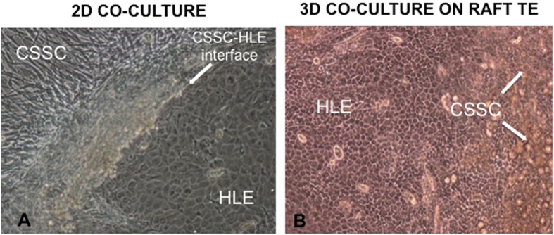
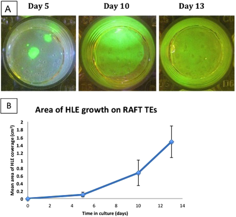
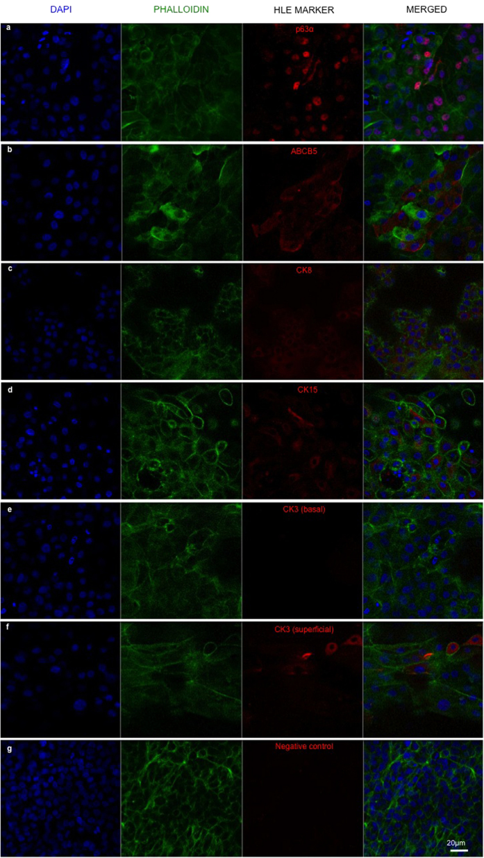
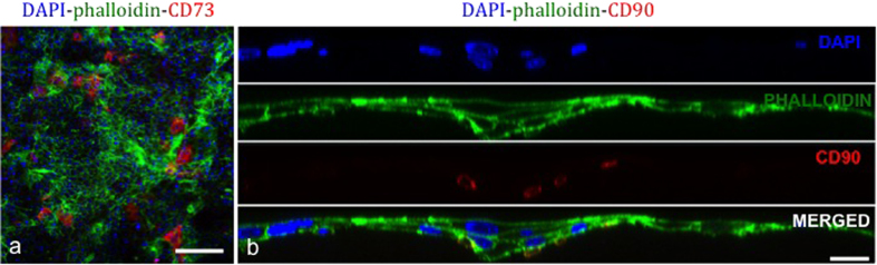
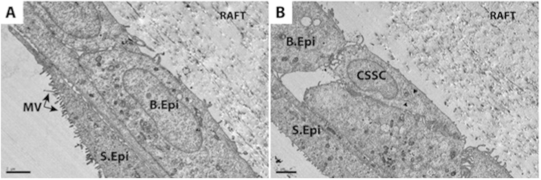
Human limbal epithelial cells (HLE) and corneal stromal stem cells (CSSC) reside in close proximity in vivo in the corneal limbal stem cell niche. However, HLE are typically cultured in vitro without supporting niche cells. Here, we re-create the cell-cell juxtaposition of the native environment in vitro, to provide a tool for investigation of epithelial-stromal cell interactions and to optimize HLE culture conditions for potential therapeutic application. RAFT (Real Architecture For 3D Tissue) tissue equivalents (TEs) were used as a 3-dimensional substrate for co-culturing HLE and CSSC. Our results demonstrate that a monolayer of HLE that maintained expression of p63α, ABCB5, CK8 and CK15 (HLE markers), formed on the surface of RAFT TEs within 13 days of culture. CSSC remained in close proximity to HLE and maintained expression of mesenchymal stem cell markers. This simple technique has a short preparation time of only 15 days with the onset of HLE layering and differentiation observed. Furthermore, co-cultivation of HLE with another niche cell type (CSSC) directly on RAFT TEs, eliminates the requirement for animal-derived feeder cells. RAFT TEs may be useful for future therapeutic delivery of multiple cell types to restore the limbal niche following ocular surface injury or disease.


Figures

Figure 1
Figure 1
(a) Light microscopy image showing a primary culture of a mixed population of human corneal stromal stem cells (CSSC) and human limbal epithelial cells (HLE) on plastic (passage 0, day 15). Spontaneous organization of the two different cell types is observed (CSSC-HLE interface). The characteristic cobblestone morphology of a HLE cluster is visible adjacent to confluent CSSC at its periphery (magnification ×10). (b) Light microscopy image showing a mixed population of human corneal stromal stem cells (CSSC) and human limbal epithelial cells (HLE) cultured on RAFT TE for 13 days. A confluent monolayer of epithelial cells with the characteristic cobblestone morphology is visible with clumps of CSSC (white arrows) shedding from the surface of RAFT TE (magnification ×10).
Figure 2
Figure 2
(a) Photographs of fluorescein diacetate (FdA)-stained RAFT TEs with mixed population of CSSC and HLE on surface. Images are taken with a camera using a yellow lens under a blue light. HLE growth (seen in green) increases over time in culture until a confluent monolayer is achieved by day 13. (b) Graph illustrating mean area of HLE growth (n = 4 donors) over 13 days of culture. RAFT TEs were stained with FdA and images taken at different time points during culture. Each point represents a mean value taken from the average of 4 biological donors (i.e. 4 RAFT TEs per donor).
Figure 3
Figure 3. Confocal split channel images of RAFT TEs cultured with a mixed population of CSSC and HLE for 13 days in CSSC media.
Positive staining of HLE markers (a) p63α, (b) ABCB5, (c) CK8 and (d) CK15 can be seen in red; (e) basal HLE do not express HLE differentiation marker, CK3; (f) CK3 expression sparsely observed in superficial HLE; (g) negative control without primary antibody. (Blue—DAPI; green—phalloidin). Scale bar 20 μm.
Figure 4
Figure 4. Confocal images of RAFT TEs cultured with a mixed population of CSSC and HLE for 13 days in CSSC media.
Positive staining of CSSC (mesenchymal markers) (a) CD73 expression of CSSC sparsely remaining on surface of RAFT TE in close proximity to HLE (scale bar 200 μm); (b) Split channel line scan showing cross-section of RAFT TE at day 13 of culture. CSSC with positive CD90 expression (seen in red) appear burrowed beneath basal HLE. (Blue - DAPI; Green - phalloidin). Scale bar 20 μm.
Figure 5
Figure 5. TEM micrographs of RAFT TEs cultured with a mixed population of CSSC and HLE for 13 days.
(a) multilayering of HLE with a basal epithelial cell (B.Epi) visible beneath a superficial epithelial (S.Epi) cell with typical microvilli (MV) features on the apical surface. (b) reveals the close interaction of a CSSC and a basal epithelial cell (B.Epi) on the basal layer. CSSC can be distinguished from HLE as it appears smaller than HLE with a more spindle morphology. Black arrows indicate presence of endoplasmic reticulum, which is more developed in CSSC and thus more visible. Scale bar 2 μm.

References

    1. Schofield R. The relationship between the spleen colony forming cell and the haemopoietic stem cell. Blood Cells 4, 7–25 (1978). - PubMed
    1. Schermer A., Galvin S. & Sun T. T. Differentiation-related expression of a major 64K corneal keratin in vivo and in culture suggests limbal location of corneal epithelial stem cells. J Cell Biol. 103(1), 49–62. (1986). - PMC - PubMed
    1. Davanger M. & Evensen A. Role of the pericorneal papillary structure in renewal of corneal epithelium. Nature 229(5286), 560–561 (1971). - PubMed
    1. Dua H. S., Joseph A., Shanmuganathan V. A. & Jones R. E. Stem cell differentiation and the effects of deficiency. Eye 17, 877–885 (2003). - PubMed
    1. Du Y., Funderburgh M. L., Mann M. M., SundarRaj N. & Funderburgh J. L. Multipotent stem cells in human corneal stroma. Stem Cells 23, 1266–1275 (2005). - PMC - PubMed

Publication types