Review
. 2015 Dec;5(12):1247-61.
doi: 10.1158/2159-8290.CD-15-0671. Epub 2015 Nov 3.

Pancreatic Cancer Metabolism: Breaking It Down to Build It Back Up

Affiliations
Review

Pancreatic Cancer Metabolism: Breaking It Down to Build It Back Up

Rushika M Perera et al. Cancer Discov. 2015 Dec.

Abstract

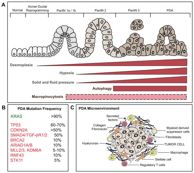
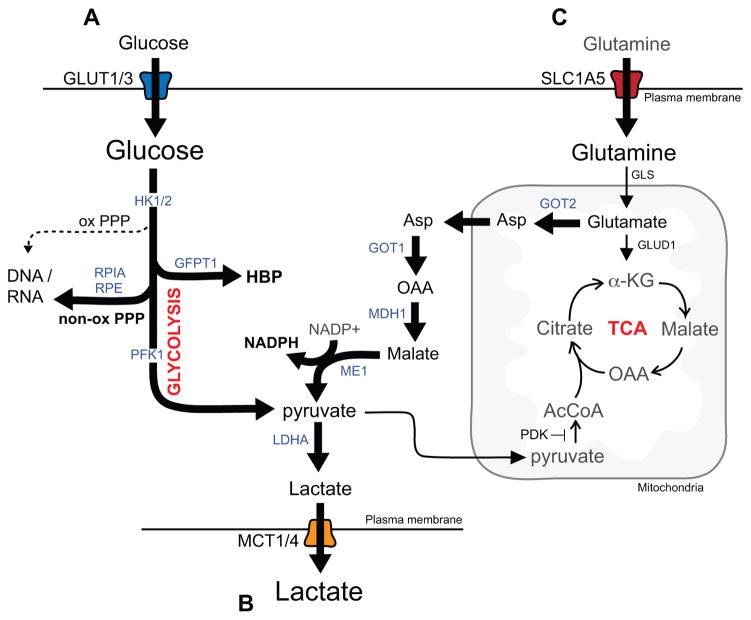
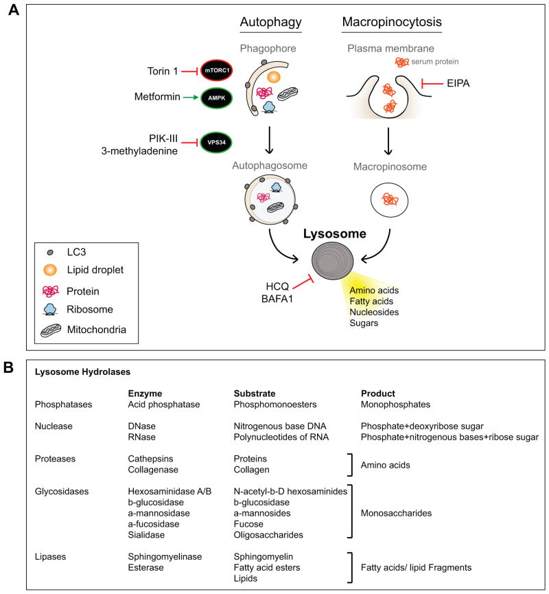
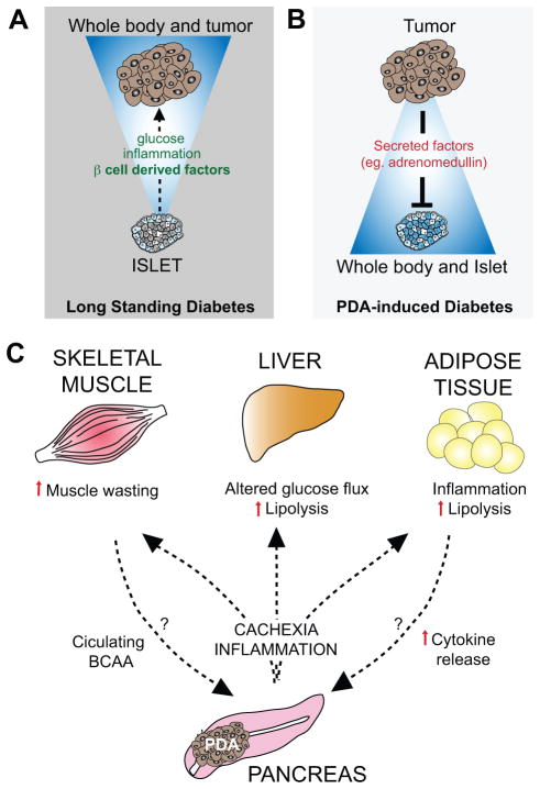
How do cancer cells escape tightly controlled regulatory circuits that link their proliferation to extracellular nutrient cues? An emerging theme in cancer biology is the hijacking of normal stress response mechanisms to enable growth even when nutrients are limiting. Pancreatic ductal adenocarcinoma (PDA) is the quintessential aggressive malignancy that thrives in nutrient-poor, hypoxic environments. PDAs overcome these limitations through appropriation of unorthodox strategies for fuel source acquisition and utilization. In addition, the interplay between evolving PDA and whole-body metabolism contributes to disease pathogenesis. Deciphering how these pathways function and integrate with one another can reveal novel angles of therapeutic attack.

Significance: Alterations in tumor cell and systemic metabolism are central to the biology of pancreatic cancer. Further investigation of these processes will provide important insights into how these tumors develop and grow, and suggest new approaches for its detection, prevention, and treatment.


Conflict of interest statement

Conflict of Interest Statement: The authors have no conflicts of interest to declare

Figures

Figure 1
Figure 1. Schematic of the multi-stage progression of pancreatic ductal adenocarcinoma
A) PDA arises from the multi-stage progression of precursor lesions known as pancreatic intraepithelial neoplasia (PanIN). B) KRAS mutations are an early event in disease pathogenesis, present in the great majority of early stage PanIN lesions. Mutations in a series of tumor suppressors occur as later events, and contribute to disease progression. C) PDA is also associated with evolving alterations in the tumor microenvironment, including increasing fibrosis and extracellular matrix deposition (desmoplasia) and recruitment of immune and inflammatory cells. Increasing desmoplasia accompanies progressive disease (as indicated) and creates intratumoral pressure that compresses the vasculature, resulting in limited blood flow to the tumor and consequent hypoxia and low nutrient delivery. In turn, PDA cells exhibit activation of nutrient scavenging pathways (autophagy and macropinocytosis) that support tumor cell growth. While autophagy activation is a late event in PDA tumorigenesis, the precise temporal dynamics of macropinocytosis is as yet unknown (dotted box).
Figure 2
Figure 2. Alterations in metabolite utilization in PDA
A) KRAS promotes glucose metabolism in PDA cells by upregulating the GLUT1 transporter and driving glycolysis through induction of the expression of multiple glycolytic enzymes. In addition, glycolytic intermediates are shunted toward biosynthetic pathways including the non-oxidative arm of the pentose phosphoate pathway (PPP) for synthesis of DNA and RNA and the hexosamine biosynthesis pathway (HBP), which generates precursors necessary for generation of glycoproteins, glycolipids, proteoglycans and glycosaminoglycans. B) In addition, PDA cells have enhanced activity of monocarboxylate transporters, MCT1 and MCT4 that shuttle lactate in order to prevent intracellular accumulation and subsequent decreases in cytosolic pH. C) KRAS also activates and reprograms glutamine metabolism. A proportion of glutamate is utilized to fuel NAPDH production via the aspartate-malate shunt thus contributing to maintenance of reduced glutathione levels and redox balance. The enzymes whose expression levels are regulated by mutant Kras are indicated in blue.
Figure 3
Figure 3. Nutrient scavenging in PDA converges at the lysosome for breakdown of intracellular and extracellular cargo
A) PDA cells show enhanced autophagy activation and macropinocytosis in vitro and in vivo. Autophagy involves formation of double membrane vesicles that surround a portion of cytoplasm thus encapsulating cargo material (protein, lipid, organelles) that is delivered to lytic organelles (lysosome) for breakdown. Positive (AMPK and VPS34) and negative (mTORC1) kinase regulators of autophagy are indicated. Macropinocytosis, the bulk uptake of extracellular material, occurs via plasma membrane invagination and generation of internalized macropinosomes. These cargo-laden vesicles similarly fuse with lysosomes for efficient degradation of the internalized material. Therefore lysosomes are a key central delivery port for substrates destined for breakdown and serve to recycle the constituent building blocks and support cellular metabolism. Drugs that modulate different aspects of these pathways are shown. B) Resident lysosomal enzymes, their substrates and final products are listed.
Figure 4
Figure 4. PDA is linked to alterations in whole body metabolism
A) Conditions associated with altered systemic metabolism—namely long standing diabetes and obesity—are associated with increased PDA risk. In the case of diabetes, the increased secretion of islet-derived factors such as insulin may make particular contributions to PDA development. B) PDAs can reciprocally induce diabetes as a paraneoplastic syndrome (referred to as PDA-induced diabetes or recent-onset diabetes) by secretion of tumor-associated factors (e.g. adrenomedullin) that cause beta cell dysfunction. C) Advanced PDA is associated with cachexia, a condition involving weight loss and altered function of several metabolic tissues (skeletal muscle, liver and adipose tissue). Cachexia is thought to be induced by inflammatory mediators and cytokines produced by the PDA cells themselves as well as components of the PDA microenvironment. In addition, increased pools of circulating branched chain amino acids (BCAA) are an early sign of PDA onset, and may also be liberated from the muscle prior to clinically evident cachexia. These BCAA and the breakdown products of muscle and adipose tissue in cachexia may in turn serve as fuel sources that feed tumor growth.

References

    1. Ryan DP, Hong TS, Bardeesy N. Pancreatic adenocarcinoma. N Engl J Med. 2014;371:2140–1. - PubMed
    1. Maitra A, Hruban RH. Pancreatic cancer. Annu Rev Pathol. 2008;3:157–88. - PMC - PubMed
    1. Morris JPt, Wang SC, Hebrok M. KRAS, Hedgehog, Wnt and the twisted developmental biology of pancreatic ductal adenocarcinoma. Nat Rev Cancer. 2010;10:683–95. - PMC - PubMed
    1. Aguirre AJ, Bardeesy N, Sinha M, Lopez L, Tuveson DA, Horner J, et al. Activated Kras and Ink4a/Arf deficiency cooperate to produce metastatic pancreatic ductal adenocarcinoma. Genes Dev. 2003;17:3112–26. - PMC - PubMed
    1. Collisson EA, Sadanandam A, Olson P, Gibb WJ, Truitt M, Gu S, et al. Subtypes of pancreatic ductal adenocarcinoma and their differing responses to therapy. Nat Med. 2011;17:500–3. - PMC - PubMed

Publication types

MeSH terms