. 2016 Mar;95(3):292-301.
doi: 10.1177/0022034515613508. Epub 2015 Nov 3.

Excess BMP Signaling in Heterotopic Cartilage Forming in Prg4-null TMJ Discs

Affiliations

Excess BMP Signaling in Heterotopic Cartilage Forming in Prg4-null TMJ Discs

T E Bechtold et al. J Dent Res. 2016 Mar.

Abstract

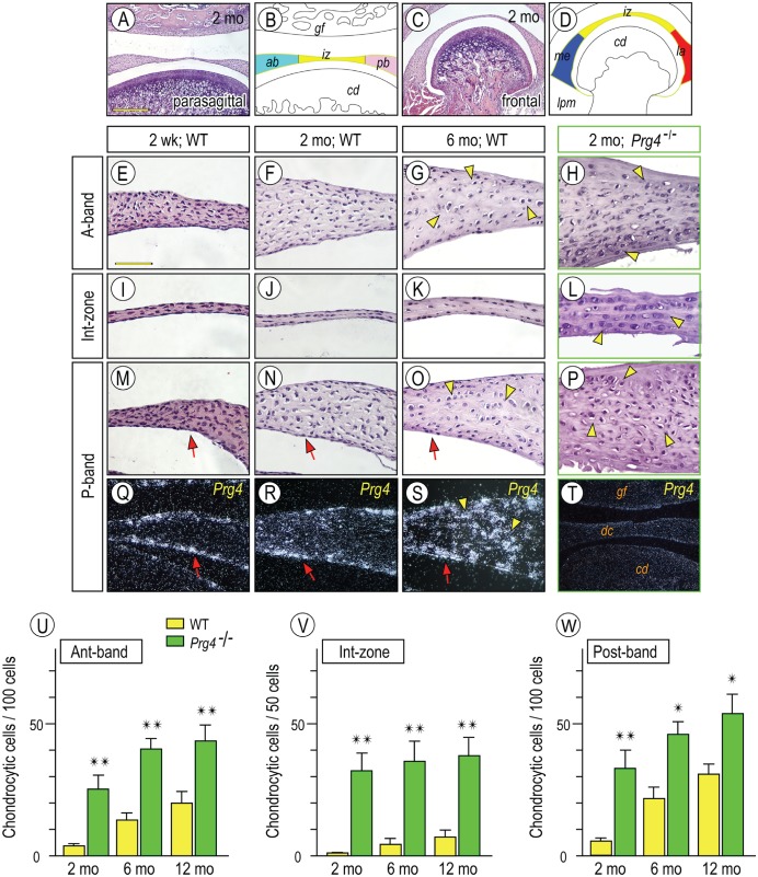
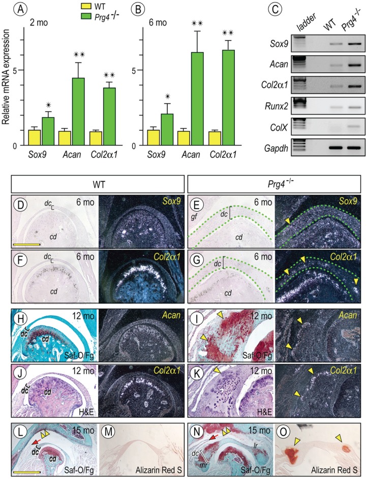
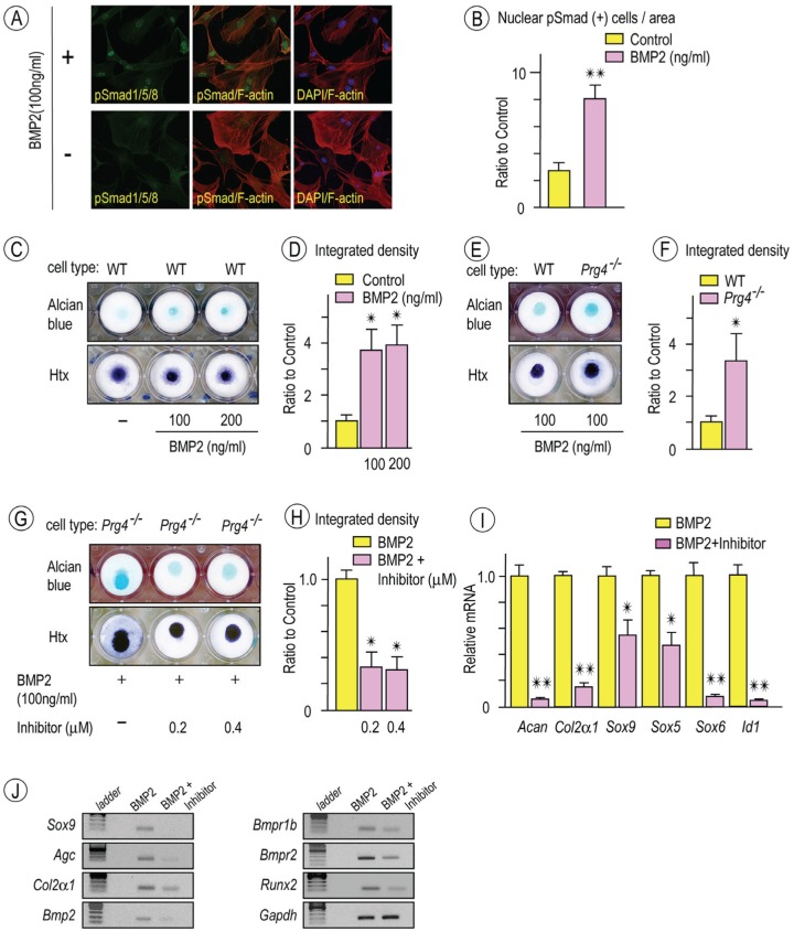
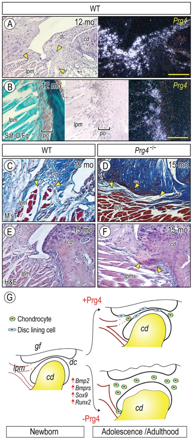
Heterotopic cartilage develops in certain pathologic conditions, including those affecting the human temporomandibular joint (TMJ), but the underlying molecular mechanisms remain obscure. This is in part due to the fact that a reliable animal model of such TMJ diseases is not available. Here, we show that aberrant chondrocyte differentiation and ectopic cartilage formation occur spontaneously in proteoglycan 4 (Prg4) mutant TMJ discs without further invasive procedure. By 2 mo of age, mutant disc cells displayed chondrocyte transdifferentiation, accompanied by strong expression of cartilage master gene Sox9 and matrix genes aggrecan and type II collagen. By 6 mo, heterotopic cartilage had formed in the discs and expressed cartilage hypertrophic markers Runx2 and ColX. The ectopic tissue grew in size over time and exhibited regional mineralization by 12 mo. Bone morphogenetic protein (BMP) signaling was activated with the ectopic chondrogenic cells and chondrocytes, as indicated by phosphorylated Smad 1/5/8 nuclear staining and by elevated expression of Bmp2, Bmpr1b, Bmpr2, and BMP signaling target genes. Likewise, we found that upon treatment with recombinant human BMP 2 in high-density micromass culture, mutant disc cells differentiated into chondrocytes and synthesized cartilage matrix more robustly than control cells. Importantly, a specific kinase inhibitor of BMP receptors drastically attenuated chondrogenesis in recombinant human BMP 2-treated mutant disc cultures. Unexpectedly, we found that Prg4 was expressed at joint-associated sites, including disc/muscle insertion and muscle/bone interface, and all these structures were abnormal in Prg4 mutants. Our data indicate that Prg4 is needed for TMJ disc integrity and function and that its absence leads to ectopic chondrogenesis and cartilage formation in conjunction with abnormal BMP signaling. Our findings imply that the BMP signaling pathway could be a potential therapeutic target for prevention or inhibition of ectopic cartilage formation in TMJ disease.

Keywords: TMJ disorder; articular disc; ectopic cartilage; joint lubrication; lubricin; temporomandibular joint.


Conflict of interest statement

The authors declare no potential conflicts of interest with respect to the authorship and/or publication of this article.

Figures

Figure 1.
Figure 1.
Temporospatial Prg4 expression in wild-type discs and aberrant differentiation of chondrocyte-like cells in Prg4-/- discs. Diagram (B, D) illustrating respective disc portions from parasagittal (A) and frontal sections (C) of temporomandibular joints (TMJs). TMJs from mice were analyzed by histology: 2-wk-old (E, I, M, Q), 2-mo-old (F, J, N, R), and 6-mo-old (G, K, O, S) control and 2-mo-old Prg4-/- (H, L, P, T). In situ hybridization with isotope-labeled riboprobes for Prg4 expression (Q–T). Note that Prg4 is expressed in disc-lining cells (arrows) and chondrocyte-like cells (arrowheads). Chondrocyte-like cells were counted in anterior (Ant) band, intermediate (Int) zone, and posterior (Post) band in distinct TMJ sections from control and Prg4-null mice at ages of 2, 6, and 12 mo (U–W; cell number is present per approximately 100 cells in anterior and posterior bands and approximately 50 cells in intermediate zone). Areas were randomly selected from 5 to 8 sections per sample (n = 3 for mouse/each group; *P < 0.05, **P < 0.01) and presented as average ± SD. Scale bars: 250 µm in A, also for C, T; 65 µm in E, also for F–S. ab, anterior band; cd, condyle; dc, disc; gf, glenoid fossa; iz, intermediate zone; la, lateral part; lpm, lateral pterygoid muscle; me, medial part; pb, posterior band; WT, wild type.
Figure 2.
Figure 2.
Increased expression of chondrocyte markers in Prg4-/- discs. Histograms depicting the relative expression of Sox9, aggrecan (Acan), and Col2α1 in Prg4-/- and control discs at 2 mo (A) and 6 mo (B) of age and presented as average ± SD (n = 3 for mouse/each group, *P < 0.05, **P < 0.01). Semiquantitative reverse transcription polymerase chain reaction reveals increased expression of early (Sox9, Acan, and Col2α1) and maturing chondrocyte (Runx2 and ColX ) markers in Prg4-/- discs as compared with control littermates at age of 6 mo (C). Frontal sections prepared from TMJs and discs from 6-mo-old (D–G), 12-mo-old (H–K), and 15-mo-old (L–O) control (D, F, H, J, L, M) and Prg4-/- (E, G, I, K, N, O) mice were processed for in situ hybridization with isotope-labeled riboprobes for Sox9 (D, E, arrowhead), Col2α1 (F, G, J, K, arrowhead), and Acan (H, I). Mutant discs were delineated by green dashed lines (E, G). Sections were also stained with safranin O (Saf-O) / fast green (Fg; H, I, L, N) and alizarin red S (M, O) and evaluate proteoglycans and/or mineralization in discs, respectively. Safranin O–stained cartilage (I, bright field, arrowheads) was composed of Col2α1- and Acan-expressing chondrocytes (I, K, dark field, arrowheads). Alizarin red S–stained mineralized tissues were detected in the mesial and lateral portions of the mutant discs (O, arrowheads). Note also the massive safranin O–stained cartilage developed in the mutant glenoid fossa as compared with control (L, N, double arrowhead, respectively) and the elongated medial synovial membrane in mutants (L, N, arrow). Scale bars: 1.2 mm in D, also for E–K, M, O; 2.5 mm in L, also for N. cd, condyle; dc, disc; gf, glenoid fossa; H&E, hematoxylin and eosin; WT, wild type.
Figure 3.
Figure 3.
Increased bone morphogenetic protein (BMP) signaling in Prg4-/- discs. Histograms depicting the relative expression of Bmp2, Bmpr1b, and Bmpr2 in Prg4-/- and control discs at age of 2 mo (A) and 6 mo (B) and presented as average ± SD (n = 3 for mouse/each group, *P < 0.05, **P < 0.01). Semiquantitative reverse transcription polymerase chain reaction showing that increased expression of Bmp2, Bmp type I and type II receptors, and Id-1 in discs from Prg4-/- mice as compared with wild type (WT) at age of 6 mo (C). TMJ discs from 4-mo-old control (D, F, I) and Prg4-/- (E, G, H, J, K) mice were analyzed by in situ hybridization with isotope-labeled riboprobes for Bmp2 (D, E) or by immunohistochemistry with phosphorylated Smad 1/5/8 (pSmad1/5/8) antibodies. Sections not treated with primary antibodies served as control (H, K). Note increased Bmp2 expression (E; arrowheads) and pSmad1/5/8-nulear translocation (G, J; arrowheads) are detected in chondrocyte-like cells in the anterior band (Ant. Band) and the intermediate zone (Int. Zone) in Prg4-/- discs. Scale bars: 90 µm in D, also for E; 55 µm in F, also for G–K.
Figure 4.
Figure 4.
Chondrogenic potential of Prg4-/- disc cells stimulated by recombinant human bone morphogenetic protein 2 (rhBMP-2) and attenuated by bone morphogenetic protein (BMP) inhibitor. Disc cells from 2-mo-old wild-type (WT) mice were plated on coverslips and grown in serum-starved Dulbecco’s modified Eagle’s medium for 12 h. Cultures were then stimulated with rhBMP2 for 30 min, fixed, and incubated with a rabbit phosphorylated Smad 1/5/8 (pSmad1/5/8) primary antibody followed by anti-rabbit Alexa Fluor 488 (green), with rhodamine phalloidin for staining the actin cytoskeleton in cells (red; A, upper panel). Culture without rhBMP2 treatment were served as controls (A, lower panel). DAPI stains were used to detect nuclei (blue). Images of pSmad1/5/8 (green) or DAPI (blue) were merged with rhodamine phalloidin-stained cytoskeletal images (red). Disc cells with pSmad1/5/8 in the nucleus were counted in randomly selected 6 to 8 areas (approximately 15 to 20 cells/area; n = 5, **P < 0.01) and presented as average ± SD (B). Day 10 micromass cultures of WT disc cells stimulated with rhBMP-2 (100 or 200 ng/mL) exhibited increased alcian blue staining compared with control cultures (C, upper panel; n = 4). Micromass cultures were also counterstained with hematoxylin (Htx) to reveal the presence of cells in cultures (C, lower panel). Integrated density of alcian blue–stained cultures was measured (D; n = 4, *P < 0.05). Day 10 micromass cultures of Prg4-/- disc cells stimulated with rhBMP-2 (100 ng/mL; E, upper panel) revealed increased alcian blue staining compared to WT disc cells (E, upper panel). Htx counterstaining (E; lower panel). Integrated density of alcian blue–stained cultures were measured (F; n = 4, *P < 0.05). Micromass cultures of Prg4-/- disc cells were treated with rhBMP-2 (100 ng/mL) alone or rhBMP-2 (100 ng/mL) / LDN-193189 (BMP inhibitor) at indicated concentration (G). Alcian blue staining (G, upper panel) and Htx counterstaining (G; lower panel). Integrated density of alcian blue–stained cultures was measured (H; n = 4, *P < 0.05). Histograms depicting relative expression of chondrocyte makers, Sox5, Sox6, Sox9, and BMP target were decreased in BMP inhibitor-treated cultures as compared with controls (I; n = 3, *P < 0.05, **P < 0.01). Semiquantitative reverse transcription polymerase chain reaction revealed decreased expression of chondrocyte markers, Bmp2, Bmp receptors, and Runx2 in Prg4-/- discs as compared with controls (J). Graphs depict mean ± SD. Control data were set as 1 (D, F, H, I).
Figure 5.
Figure 5.
Prg4 expression at nonarticulation sites and tissue derangement in Prg4-/- mice. Parasagittal sections of TMJs from 12-mo (A, B) and 15-mo (C–F) control (A, B, C, E) and Prg4-/- (D, F) mice were processed for histology and in situ hybridization with isotope-labeled riboprobes for Prg4 expression (A, B). Sections were stained with safranin O/fast green (Saf-O/Fg; B), Masson’s trichrome (M’sT; C, D), and hematoxylin and eosin (H&E; E, F). Note that Prg4 is expressed at disc/lateral pterygoid muscle junction (A, arrowheads) and muscle/tendon insertion site into the mandibular condyle (B). Note also the disrupted muscle insertion into the articular disc in mutant (D, arrowheads) as compared with control (C, arrowheads) and ectopic cartilage formation at muscle/tendon insertion site into the mandibular condyle in mutant (F, arrowheads). (G) Model of heterotopic ossification in TMJs caused by absence of Prg4 action and involvement of abnormal BMP signaling. (G) Model of increased chondrogenesis and subsequent cartilage formation in Prg4 mutant discs. TMJ appears to develop normally, and the disc separates the joint space into the upper and lower joint cavities. With further development, the disc displays a concaved shape; the disc-lining cells cover the surface of the disc; and a few chondrogenic cells and chondrocytes differentiate in the anterior and posterior bands in wild-type disc (with Prg4 action). In sharp contrast, in Prg4 mutant mice (without Prg4 action), the disc becomes much thicker, and this is accompanied by increased chondrocyte number and ectopic cartilage formation throughout the disc. In addition, the lateral pterigoid muscle loses its smooth integration into the disc. The increased chondrogenesis and subsequent cartilage formation throughout the disc are accompanied by activation of Bmp signaling and increased expression of Sox9 and Runx2. Scale bars: 120 µm in A, also for E, F; 60 µm in B, also for C, D. cd, condyle; dc, disc; lpm, lateral pterygoid muscle; po, periosteum.

References

    1. Asai S, Otsuru S, Candela ME, Cantley L, Uchibe K, Hofmann TJ, Zhang K, Wapner KL, Soslowsky LJ, Horwitz EM, et al. 2014. Tendon progenitor cells in injured tendons have strong chondrogenic potential: the CD105-negative subpopulation induces chondrogenic degeneration. Stem Cells. 32(12):3266–3277. - PMC - PubMed
    1. Bahabri SA, Suwairi WM, Laxer RM, Polinkovsky A, Dalaan AA, Warman ML. 1998. The camptodactyly-arthropathy-coxa vara-pericarditis syndrome: clinical features and genetic mapping to human chromosome 1. Arthritis Rheum. 41(4):730–735. - PubMed
    1. Berkovitz BK, Pacy J. 2000. Age changes in the cells of the intra-articular disc of the temporomandibular joints of rats and marmosets. Arch Oral Biol. 45(11):987–995. - PubMed
    1. Billings PC, Fiori JL, Bentwood JL, O’Connell MP, Jiao X, Nussbaum B, Caron RJ, Shore EM, Kaplan FS. 2008. Dysregulated BMP signaling and enhanced osteogenic differentiation of connective tissue progenitor cells from patients with fibrodysplasia ossificans progressiva (FOP). J Bone Miner Res. 23(3):305–313. - PMC - PubMed
    1. Chen D, Ji X, Harris MA, Feng JQ, Karsenty G, Celeste AJ, Rosen V, Mundy GR, Harris SE. 1998. Differential roles for bone morphogenetic protein (BMP) receptor type IB and IA in differentiation and specification of mesenchymal precursor cells to osteoblast and adipocyte lineages. J Cell Biol. 142(1):295–305. - PMC - PubMed

Publication types

MeSH terms

Substances

LinkOut - more resources