Review
. 2015 Nov 1;142(21):3620-9.
doi: 10.1242/dev.120394.

Progress and renewal in gustation: new insights into taste bud development

Affiliations
Review

Progress and renewal in gustation: new insights into taste bud development

Linda A Barlow. Development. .

Abstract

The sense of taste, or gustation, is mediated by taste buds, which are housed in specialized taste papillae found in a stereotyped pattern on the surface of the tongue. Each bud, regardless of its location, is a collection of ∼100 cells that belong to at least five different functional classes, which transduce sweet, bitter, salt, sour and umami (the taste of glutamate) signals. Taste receptor cells harbor functional similarities to neurons but, like epithelial cells, are rapidly and continuously renewed throughout adult life. Here, I review recent advances in our understanding of how the pattern of taste buds is established in embryos and discuss the cellular and molecular mechanisms governing taste cell turnover. I also highlight how these findings aid our understanding of how and why many cancer therapies result in taste dysfunction.

Keywords: Ageusia; Cell lineage; Chemotherapy; Dysgeusia; Gustatory system; Lingual organoids; Molecular genetics; Sonic hedgehog; Stem cells; Taste buds; Taste dysfunction; Wnt/β-catenin.


Conflict of interest statement

Competing interests

The author declares no competing or financial interests.

Figures

Fig. 1.
Fig. 1.
The locations of taste papillae and taste buds in the rodent tongue. Lingual taste buds are housed in distributed fungiform papillae (FFP; blue) in the anterior region of the tongue, which is otherwise covered with mechanosensory filiform papillae (flp in lower inset). Bilateral foliate papillae (FolP; blue) and a single midline circumvallate papilla (CVP; blue) are located posteriorly in the tongue. Each FFP houses one taste bud, whereas the CVP and FolP house several hundred taste buds each (depicted for the CVP only). The CVP comprises two epithelial trenches that extend ventrally from the tongue surface (asterisks in upper inset), and taste buds are aligned orthogonal to the trench axes and embedded in both medial (m) and lateral (l) trench epithelia. D, dorsal; V, ventral; A, anterior; P, posterior; R, animal's right; L, animal's left.
Fig. 2.
Fig. 2.
The sequence of taste bud and papilla development in mouse embryos. (A) At E11.5, the tongue rudiment is covered by a homogeneous epithelial bilayer, with basal and superficial keratinocytes, which is immediately above the subepithelial mesenchyme. (B) At E12.5, taste placodes form within the epithelium as clusters of columnar cells. Sensory nerve fibers arrive at taste placodes, but do not penetrate the placode epithelium until E15.5. (C,D) Taste papilla morphogenesis begins at ∼E15.5 (C), as the epithelium invaginates into the mesenchyme to form mushroom-shaped papillae with defined mesenchymal cores and immature taste buds situated apically by E18.5 (D). (E) Within a week of birth (here at P4), most FFP house differentiated taste buds. Mouse gestation is 18-19 days.
Fig. 3.
Fig. 3.
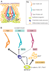
Taste cell renewal in adult mice. (A) The cell types found in adult taste buds. Basal keratinocytes (green) divide asymmetrically to self-renew (curved arrows) and generate daughter cells (straight arrows), which become non-taste cells (differentiated keratinocytes) or contribute to taste buds. Newly generated taste cells are oval shaped and express Shh (purple). Shh+ cells are postmitotic and differentiate into each of the three taste cell types (tripartite arrows). Fifty percent of Shh+ cells become Type I cells (thick arrow), while 20-30% differentiate as Type II (intermediate arrow) and <10% as Type III (thin arrow) (Miura et al., 2014). (B) Model of molecular regulation of taste cell renewal (based on Castillo et al., 2014; Gaillard et al., 2015). K5+/K14+ taste progenitor cells require β-catenin, and different levels of β-catenin within progenitors dictate daughter cell fate. Low β-catenin promotes K13+ keratinocytes, whereas higher levels allow daughter cells to acquire a taste fate, i.e. to become Shh+ taste cell precursors, which in turn differentiate into Type I, II or III cells in response to high, mid or low β-catenin, respectively. Shh signaling to K5+/K14+ progenitors promotes differentiation of all taste cell types.

References

    1. Álvarez-Buylla A. and Ihrie R. A. (2014). Sonic hedgehog signaling in the postnatal brain. Semin. Cell Dev. Biol. 33, 105-111. 10.1016/j.semcdb.2014.05.008 - DOI - PMC - PubMed
    1. Arnold K., Sarkar A., Yram M. A., Polo J. M., Bronson R., Sengupta S., Seandel M., Geijsen N. and Hochedlinger K. (2011). Sox2+ adult stem and progenitor cells are important for tissue regeneration and survival of mice. Cell Stem Cell 9, 317-329. 10.1016/j.stem.2011.09.001 - DOI - PMC - PubMed
    1. Atema J. (1971). Structures and functions of the sense of taste in the catfish (Ictalurus natalis). Brain Behav. Evol. 4, 273-294. 10.1159/000125438 - DOI - PubMed
    1. Atwood S. X., Whitson R. J. and Oro A. E. (2014). Advanced treatment for basal cell carcinomas. Cold Spring Harb. Perspect. Med. 4, a013581 10.1101/cshperspect.a013581 - DOI - PMC - PubMed
    1. Avilés E. C., Wilson N. H. and Stoeckli E. T. (2013). Sonic hedgehog and Wnt: antagonists in morphogenesis but collaborators in axon guidance. Front. Cell. Neurosci. 7, 86 10.3389/fncel.2013.00086 - DOI - PMC - PubMed

Publication types

LinkOut - more resources