. 2015 Nov 1;142(21):3637-48.
doi: 10.1242/dev.126011.

Inactivating the permanent neonatal diabetes gene Mnx1 switches insulin-producing β-cells to a δ-like fate and reveals a facultative proliferative capacity in aged β-cells

Affiliations

Inactivating the permanent neonatal diabetes gene Mnx1 switches insulin-producing β-cells to a δ-like fate and reveals a facultative proliferative capacity in aged β-cells

Fong Cheng Pan et al. Development. .

Abstract

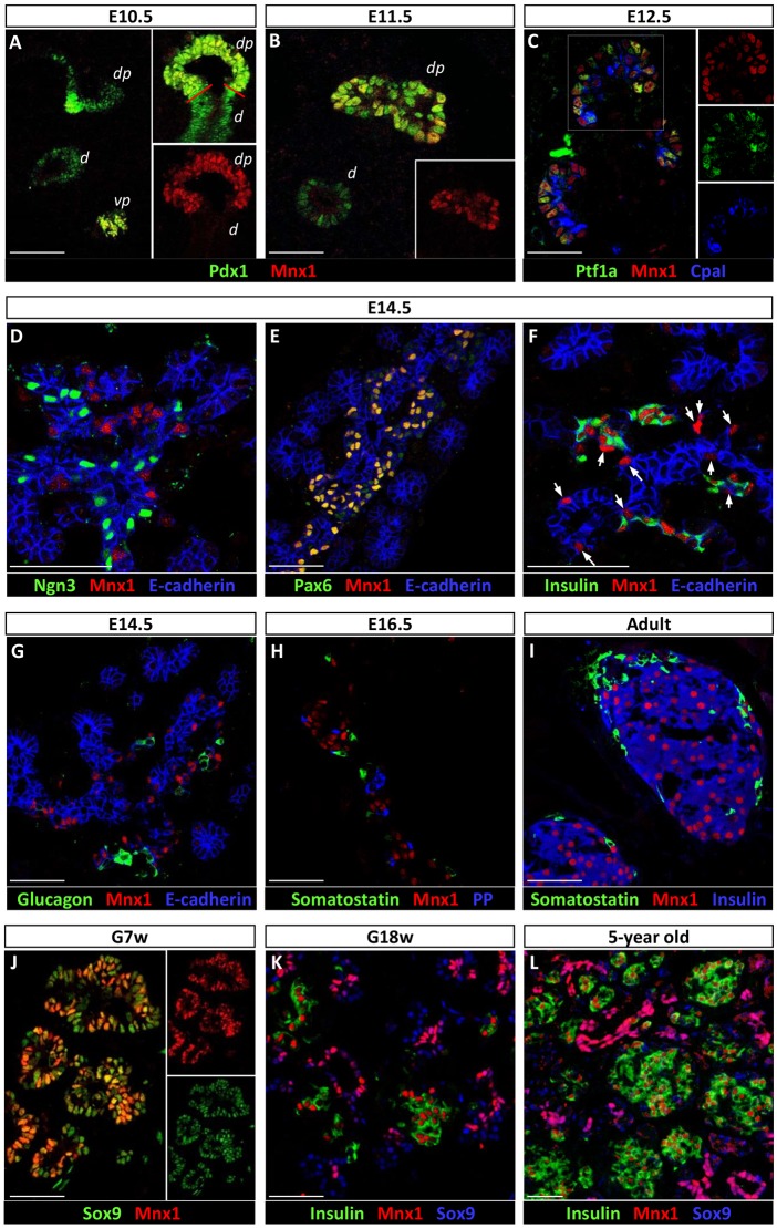
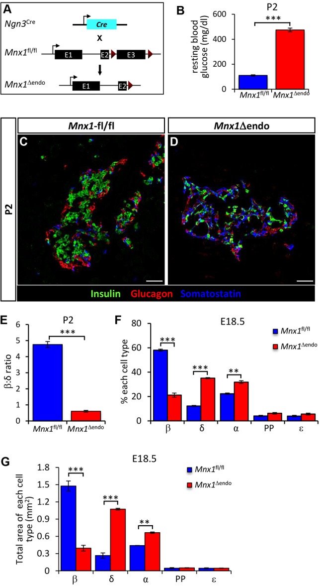
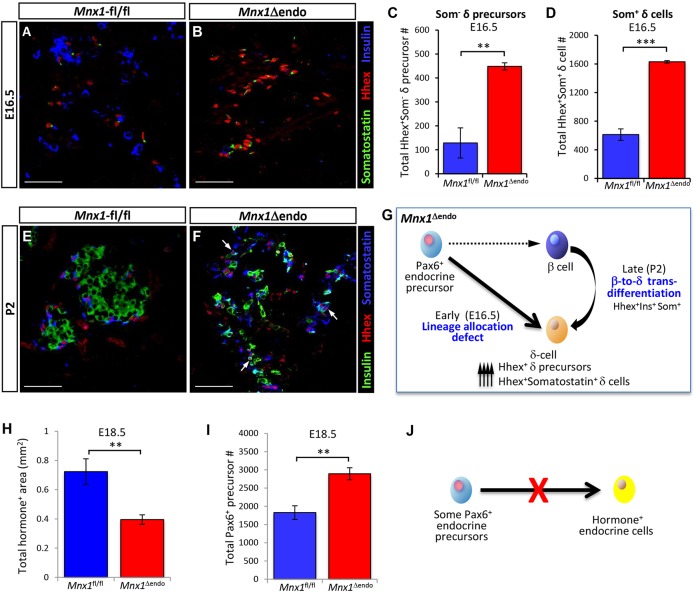
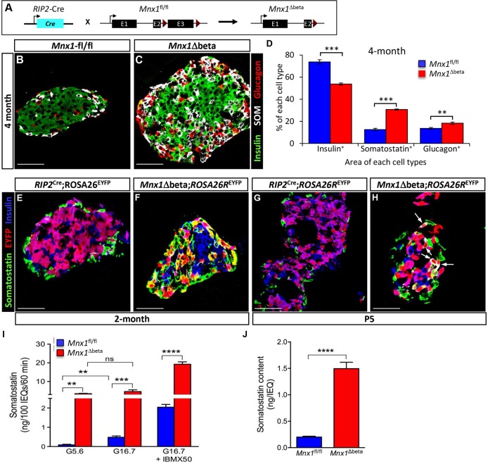
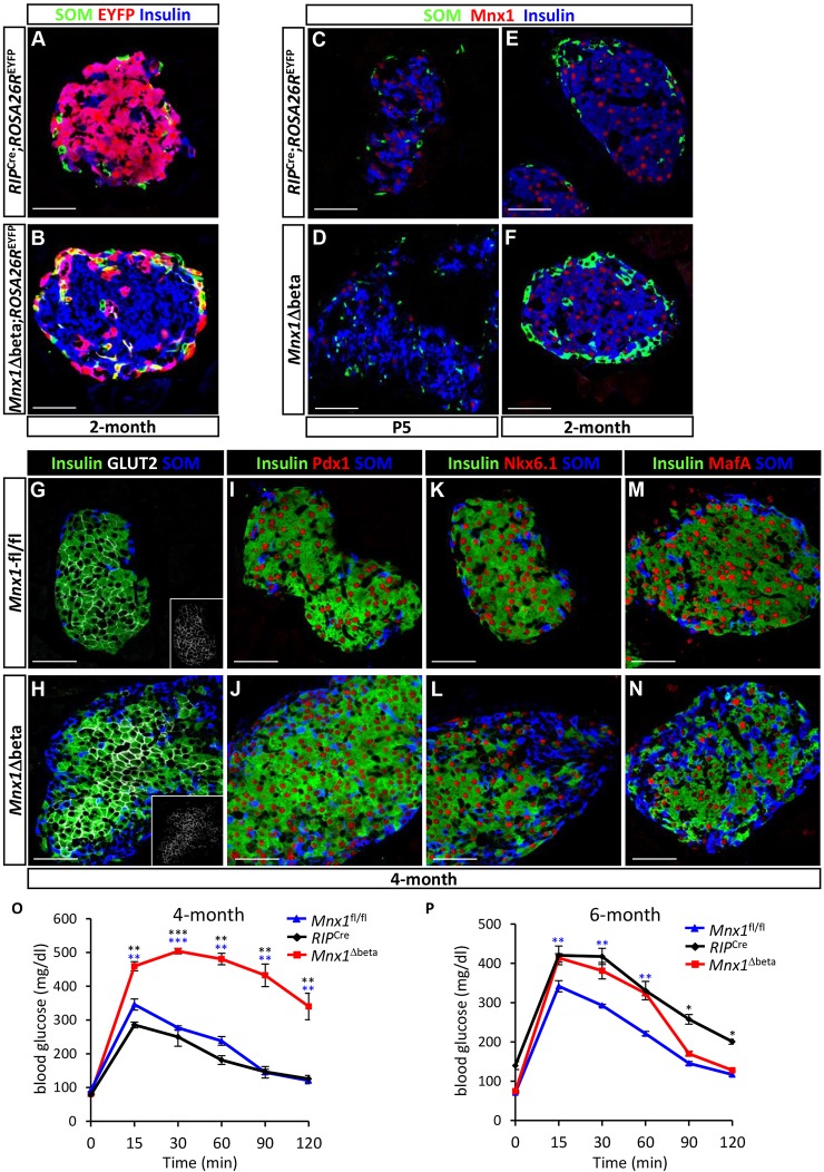
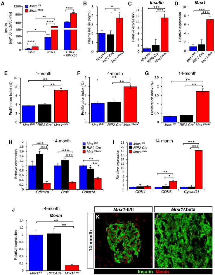
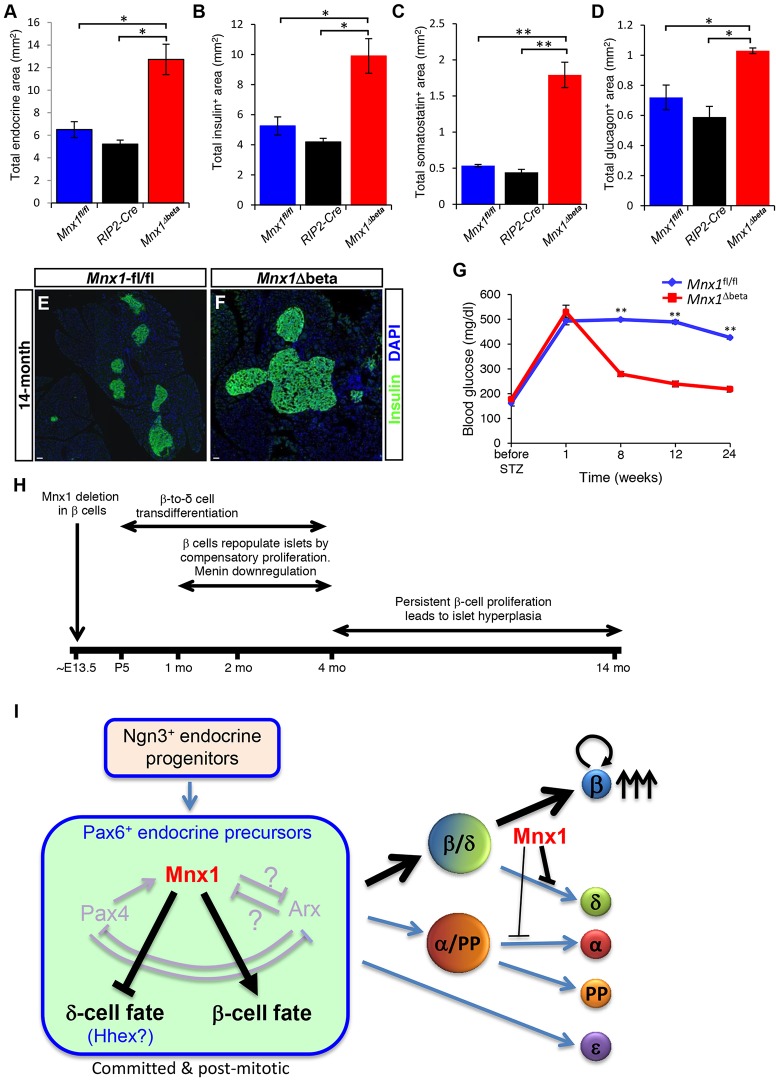
Homozygous Mnx1 mutation causes permanent neonatal diabetes in humans, but via unknown mechanisms. Our systematic and longitudinal analysis of Mnx1 function during murine pancreas organogenesis and into the adult uncovered novel stage-specific roles for Mnx1 in endocrine lineage allocation and β-cell fate maintenance. Inactivation in the endocrine-progenitor stage shows that Mnx1 promotes β-cell while suppressing δ-cell differentiation programs, and is crucial for postnatal β-cell fate maintenance. Inactivating Mnx1 in embryonic β-cells (Mnx1(Δbeta)) caused β-to-δ-like cell transdifferentiation, which was delayed until postnatal stages. In the latter context, β-cells escaping Mnx1 inactivation unexpectedly upregulated Mnx1 expression and underwent an age-independent persistent proliferation. Escaper β-cells restored, but then eventually surpassed, the normal pancreatic β-cell mass, leading to islet hyperplasia in aged mice. In vitro analysis of islets isolated from Mnx1(Δbeta) mice showed higher insulin secretory activity and greater insulin mRNA content than in wild-type islets. Mnx1(Δbeta) mice also showed a much faster return to euglycemia after β-cell ablation, suggesting that the new β-cells derived from the escaper population are functional. Our findings identify Mnx1 as an important factor in β-cell differentiation and proliferation, with the potential for targeting to increase the number of endogenous β-cells for diabetes therapy.

Keywords: Endocrine lineage diversification; Islet; β-cell fate maintenance; β-cell proliferation; β-cell versus δ-cell fate selection.


Conflict of interest statement

Competing interests

The authors declare no competing or financial interests.

Figures

Fig. 1.
Fig. 1.
Immunodetection of Mnx1 in developing and adult mouse and human pancreas. (A,B) Immunolabeling comparison of Pdx1 with Mnx1 in the embryonic mouse pancreas at E10.5 (A) and E11.5 (B). (C) Mnx1 detected in Ptf1a+ Cpa1+ tip MPC at E12.5. (D) Mnx1 is absent in Ngn3+ endocrine progenitors, but present in Pax6+ endocrine precursors (E). (F) Mnx1 can be found in insulin+ developing β-cells as early as E14.5. Arrows indicate Mnx1 expression in insulin-endocrine precursors. (G,H) Mnx1 protein was not detected in α-cells (G), δ-cells or in PP cells (H). (I) Mnx1 is restricted to β-cells in adult mouse pancreas. (J) In human fetal pancreas, Mnx1 is detected in the Sox9+ MPC population at gestational week 7 (G7w). (K,L) In G18w (K) and 5-year-old tissue (L), Mnx1 was immunodetected in β-cells and Sox9+ trunk/duct cells. Scale bars: 50 µm.
Fig. 2.
Fig. 2.
Dramatic increase in δ-cell numbers and significant decrease in β-cell numbers in Mnx1Δendo. (A) Schematic of conditional Mnx1 deletion in endocrine progenitors with Ngn3-Cre. (B) Random blood-glucose measurement showed that Mnx1Δendo mutant pups were hyperglycemic at P2 (n=10). (C,D) Immunolabeling with insulin, glucagon and somatostatin showed a dramatic increase in δ-cell number, concomitant with a decrease β-cell number at P2. (E) Significant decrease in β:δ cell ratio in Mnx1Δendo mutants at P2 (n=4). (F,G) Quantification of endocrine cell types fraction (F) and area (G) at E18.5, indicating significant increase in δ-cells at the expense of β-cells in Mnx1Δendo pancreas (n=4). Scale bars: 50 µm. Data shown as mean±s.e.m. *P<0.05, **P<0.005, ***P<0.001.
Fig. 3.
Fig. 3.
Embryonic endocrine lineage-allocation defect, followed by postnatal β-to-δ transdifferentiation as two major routes to increased δ-cell numbers. (A,B) Representative sections from E16.5 pancreata with immunodetection of Hhex, insulin and somatostatin illustrate the increase in Hhex+ somatostatin δ-cell precursors in Mnx1Δendo mutants. (C,D) Quantitative analysis of E16.5 Mnx1fl/fl and Mnx1Δendo pancreata indicate a dramatic increase in total number Hhex+somatostatin δ-cell precursors (C) and total Hhex+somatostatin+ δ-cell numbers (D) in Mnx1Δendo mutant. (E,F) Immunofluorescence analysis of Hhex, insulin and somatostatin on P2 pancreas demonstrates the presence of Hhex+insulin+somatostatin+ cells (white arrows) in Mnx1Δendo pancreas tissue, an indirect indicator of β-to-δ transdifferentiation. (G) Model showing early stage endocrine lineage-allocation defects and late-stage β-to-δ transdifferentiation when Mnx1 is inactivated in endocrine progenitors. (H-J) Quantification indicates that total endocrine area was reduced in Mnx1Δendo mutant (H), but total Pax6+ endocrine precursors were increased in Mnx1Δendo mutant at E18.5 (I), indicating failure of a subset of Pax6+ precursors to differentiate towards hormone-producing endocrine cells in the absence of Mnx1 (J). Scale bars: 50 µm. Data are shown as mean±s.e.m. **P<0.005, ***P<0.001.
Fig. 4.
Fig. 4.
β-to-δ cell transdifferentiation in Mnx1Δbeta mutants. (A) Schematic showing β-cell-specific Mnx1 deletion using RIP2-Cre transgenics. (B,C) Immunolabeling with insulin, somatostatin and glucagon showed an increase in δ- and α-cells, as well as larger islet size in the Mnx1Δbeta islets. (D) Quantitative analysis of β-, δ- and α-cell fraction indicate an increased in δ- and α-cell populations concomitant with decreased in percentage of β-cells (n=3). (E,F) Lineage-tracing analysis using ROSA26REYFP reporter show co-expression of EYFP and somatostatin, indicating that a subset of δ-cells in the Mnx1Δbeta mutant are derived from β-cells. (G,H) β-to-δ transdifferentiation, as indicated by EYFP+somatostatin+insulin+ cells (arrows), can be detected as early as P5. (I,J) Measurement of islet somatostatin secretion (I) and total somatostatin content (J) in vitro indicate that mutant δ-cells hypersecrete somatostatin and actively synthesize more somatostatin compared with control islets (n=3). Somatostatin secretion was normalized to islet number. Scale bars: 50 µm. Data are shown as mean±s.e.m. **P<0.005, ***P<0.001, ****P<0.0001.
Fig. 5.
Fig. 5.
β-cells escaping Mnx1 deletion in Mnx1Δbeta repopulate the islets. (A,B) Lineage-tracing analysis using the ROSA26REYFP reporter shows that the majority of Mnx1Δbeta β-cells at 2 months of age are escaper β-cells, as they are EYFP compared with RIP2-Cre;ROSA26REYFP. (C,D) Immunolabeling with Mnx1, insulin and somatostatin show that the majority of the Mnx1Δbeta insulin+ β-cells are devoid of Mnx1 at P5. (E,F) Most of the Mnx1Δbeta insulin+ β-cells are Mnx1+. (G-N) The escaper β-cells in Mnx1Δbeta islets are Glut2+ (G,H), Pdx1+ (I,J), Nkx6.1+ (K,L) and MafA+ (M,N). (O,P) Intraperitoneal glucose tolerance tests indicate that Mnx1Δbeta mice are glucose intolerant at 4 months (O), but glucose clearance improved by 6 months of age (P). Scale bars: 50 µm. Data are shown as mean±s.e.m. *P<0.05, **P<0.005, ***P<0.001.
Fig. 6.
Fig. 6.
Escaper β-cells secrete and produce more insulin, and continue to proliferate in Mnx1Δbeta aged mice via downregulation of menin. (A,B) Measurement of insulin secretion from isolated islets (A) and plasma insulin levels (B) indicate that Mnx1Δbeta β-cells secrete more insulin (n=3). Insulin secretion was normalized to islet number. (C,D) qRT-PCR analysis indicates significantly increased mRNA expression for Ins2 (C) and Mnx1 (D) in Mnx1Δbeta β-cells (n=4). (E-G) Mnx1Δbeta β-cells showed increased proliferation as determined by Ki67 at 1 month (n=4; E), 4 months (n=4; F) and 14 months (n=4; G). (H,I) qRT-PCR analysis show decreased expression of cell-cycle inhibitors Cdkn2a, Bmi1 and Cdkn1a (H), and increased expression of cell-cycle-positive regulators Cdk6 and Ccnd1 (I). (J) qRT-PCR analysis on 4-month-old islets RNA shows that menin mRNA expression was also downregulated in the Mnx1Δbeta islets. (K,L) Immunolabeling of menin showed that menin protein was significantly reduced in Mnx1Δbeta β-cells at 14 months. Menin protein level was similar in Mnx1fl/fl and RIP2-Cre β-cells, indicating no effect on Menin from the transgenic Cre driver. Scale bars: 50 µm. Data are shown as mean±s.e.m. *P<0.05, **P<0.005, ***P<0.001, ****P<0.0001.
Fig. 7.
Fig. 7.
Sustained proliferation of escaper β-cells leads to islet hyperplasia in Mnx1Δbeta aged mice, and alleviates hyperglycemia following STZ treatment. (A-D) Quantification of total area of hormone+ (A), insulin+ (B), somatostatin+ (C) and glucagon+ (D) area demonstrate a twofold increase in total endocrine, insulin+ and glucagon+ area, in addition to the approximately fourfold increase in somatostatin+ area. (E,F) Immunolabeling with insulin shows the presence of hyperplastic islet in Mnx1Δbeta pancreata. (G) Measurement of resting blood glucose over 6-month period upon streptozotocin (STZ) treatment showed the return of blood glucose to normal levels 2 months post-STZ treatment compared with Mnx1fl/fl mice. (H) Summary of processes occurring in Mnx1Δbeta mutant. (I) Model showing novel Mnx1 function in promoting β-cell fate and suppressing δ-cell fate in the Pax6+ endocrine precursors. Mnx1 is continuously required to maintain β-cell fate in the developing β-cells. When Mnx1 β-cells transdifferentiate into δ-cells, a subset of Mnx1+ β-cells start to repopulate islets by proliferation, leading to islet hyperplasia in aged mice. Scale bars: 100 µm. Data are shown as mean±s.e.m. *P<0.05, **P<0.005.

References

    1. Alberti K. G. M. M., Christensen N. J., Christensen S. E., Hansen A. A. P., Iversen J., Lundbaek K., Seyer-Hansen K. and Orskov H. (1973). Inhibition of insulin secretion by somatostatin. Lancet 302, 1299-1301. 10.1016/S0140-6736(73)92873-0 - DOI - PubMed
    1. Bonnefond A., Vaillant E., Philippe J., Skrobek B., Lobbens S., Yengo L., Huyvaert M., Cavé H., Busiah K., Scharfmann R. et al. (2013). Transcription factor gene MNX1 is a novel cause of permanent neonatal diabetes in a consanguineous family. Diabetes Metab. 39, 276-280. 10.1016/j.diabet.2013.02.007 - DOI - PubMed
    1. Brissova M., Aamodt K., Brahmachary P., Prasad N., Hong J.-Y., Dai C., Mellati M., Shostak A., Poffenberger G., Aramandla R. et al. (2014). Islet microenvironment, modulated by vascular endothelial growth factor-A signaling, promotes β cell regeneration. Cell Metab. 19, 498-511. 10.1016/j.cmet.2014.02.001 - DOI - PMC - PubMed
    1. Broihier H. T. and Skeath J. B. (2002). Drosophila homeodomain protein dHb9 directs neuronal fate via crossrepressive and cell-nonautonomous mechanisms. Neuron 35, 39-50. 10.1016/S0896-6273(02)00743-2 - DOI - PubMed
    1. Chera S., Baronnier D., Ghila L., Cigliola V., Jensen J. N., Gu G., Furuyama K., Thorel F., Gribble F. M., Reimann F. and Herrera P. L. (2014). Diabetes recovery by age-dependent conversion of pancreatic δ-cells into insulin producers. Nature 514, 503-507. - PMC - PubMed

Publication types

MeSH terms

Supplementary concepts