

Glenoid Bone Loss Measurement in Recurrent Shoulder Dislocation: Assessment of Measurement Agreement Between CT and MRI
- PMID: 26535360
- PMCID: PMC4555629
- DOI: 10.1177/2325967114549541
Glenoid Bone Loss Measurement in Recurrent Shoulder Dislocation: Assessment of Measurement Agreement Between CT and MRI
Abstract
Background: Shoulder instability can cause both soft tissue injury and bone defects, requiring both computed tomography (CT) and magnetic resonance imaging (MRI) for a thorough workup, which results in high patient costs and radiation exposure. Prior studies in cadaveric and nonclinical models have shown promise in assessing preoperative bone loss utilizing MRI.
Purpose: To evaluate the utility of MRI in detecting and evaluating glenoid bone defects in a clinical setting. The aim was to establish whether similar information could be determined by utilizing MRI and CT in a population with recurrent instability.
Study design: Cohort study (diagnosis); Level of evidence, 2.
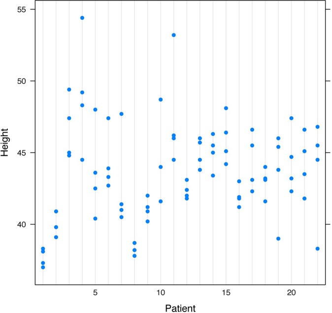
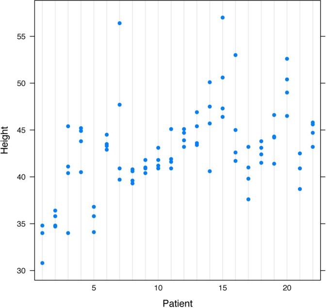
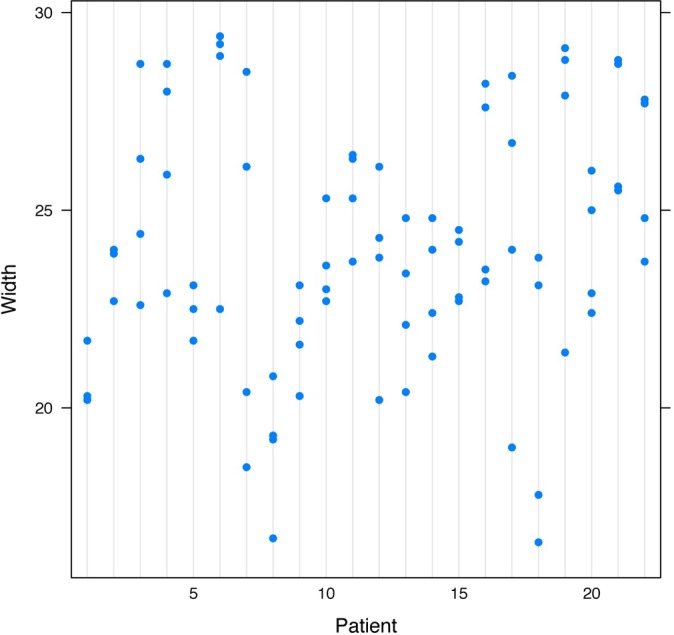
Methods: CT and MRI scans of 22 shoulders were read by 4 orthopaedic surgeons. The CT images were obtained on a 2-dimensional CT scanner. Vertical measurements were taken from the superior glenoid tubercle and directed inferiorly along the glenoid; horizontal measurements were taken across the widest part of the face of the glenoid and were perpendicular within one-half of 1° to the vertical measurement. The same protocol was followed for MRI measurements. An intraclass correlation coefficient (ICC) was calculated.
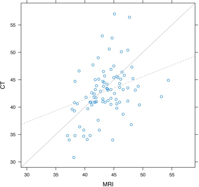
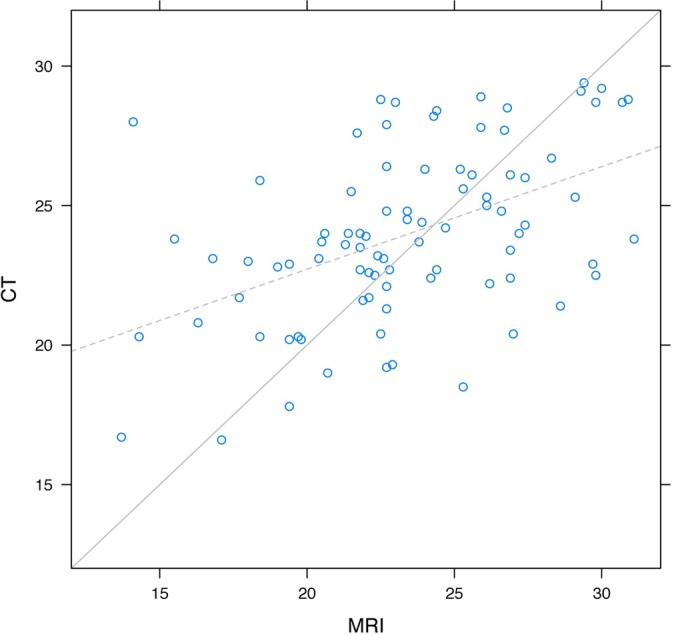
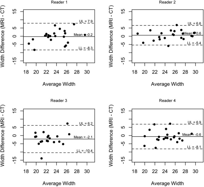
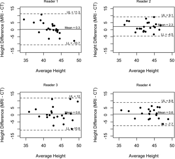
Results: There was a moderate amount of agreement between examiners for the height measurements on MRI (ICC, 0.53) and a substantial agreement for the CT images (ICC, 0.64). The width measurements for MRI had a moderate amount of agreement (ICC, 0.41), while the CT images had a fair agreement (ICC, 0.39). The height measurements between the measurements of MRI and CT images had an overall ICC of 0.43, while the width measurements had an overall ICC of 0.41, both of which were considered a moderate amount of agreement.
Conclusion: There is moderate correlation between MRI and CT scans when measuring the glenoid, indicating that taking the length-to-height ratio measurements across the glenoid is a promising way to estimate the glenoid defect. At present, a complete workup of a patient with shoulder instability includes both a CT scan and an MRI. Future research that establishes precisely how MRI misestimates CT measurements of the glenoid can perhaps obviate the need for 2 scans.
Keywords: CT; MRI; dislocation; glenoid bone loss; shoulder instability.
Conflict of interest statement
The authors declared that they have no conflicts of interest in the authorship and publication of this contribution.
Figures


References
-
- Bois AJ, Fening SD, Polster J, Jones MH, Miniaci A. Quantifying glenoid bone loss in anterior shoulder instability: reliability and accuracy of 2-dimensional and 3-dimensional computed tomography measurement techniques. Am J Sports Med. 2012;40:2569–2577. - PubMed
-
- Bushnell BD, Creighton RA, Herring MM. Bony instability of the shoulder. Arthroscopy. 2008;24:1061–1073. - PubMed
-
- Chandnani VP, Yeager TD, DeBerardino T, et al. Glenoid labral tears: Prospective evaluation with MRI imaging, MR arthrography, and CT arthrography. AJR Am J Roentgenol. 1993;161:1229–1235. - PubMed
-
- Dodson CC, Cordasco FA. Anterior glenohumeral joint dislocations. Orthop Clin North Am. 2008;39:507–518. - PubMed
LinkOut - more resources
Full Text Sources
Other Literature Sources
