. 2015 Nov 4;10(11):e0141988.
doi: 10.1371/journal.pone.0141988. eCollection 2015.

(E)-4-(3,4-Dimethoxyphenyl)but-3-en-1-ol Enhances Melanogenesis through Increasing Upstream Stimulating Factor-1-Mediated Tyrosinase Expression

Affiliations

(E)-4-(3,4-Dimethoxyphenyl)but-3-en-1-ol Enhances Melanogenesis through Increasing Upstream Stimulating Factor-1-Mediated Tyrosinase Expression

Jisu Park et al. PLoS One. .

Abstract

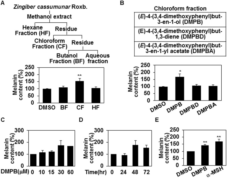
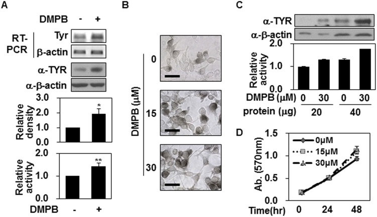
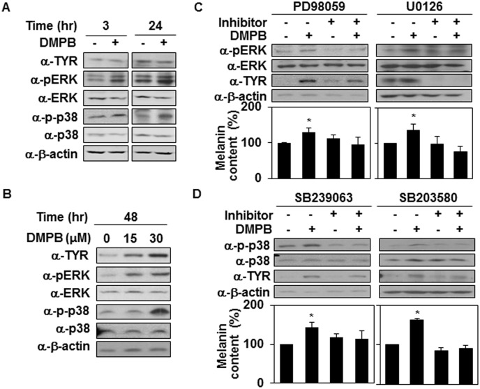
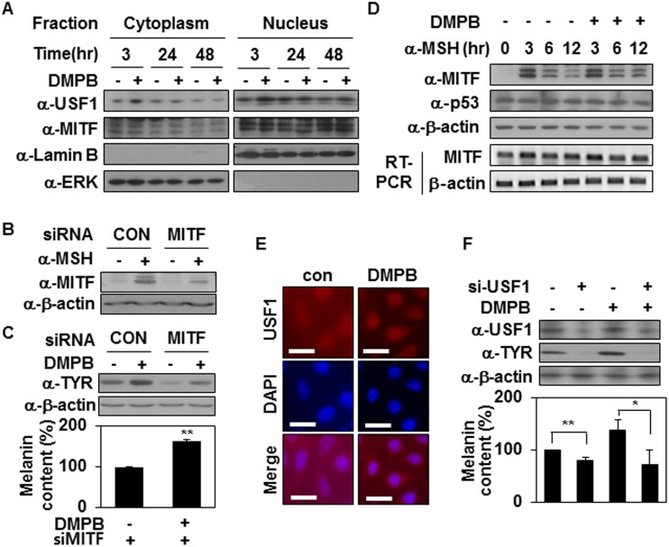
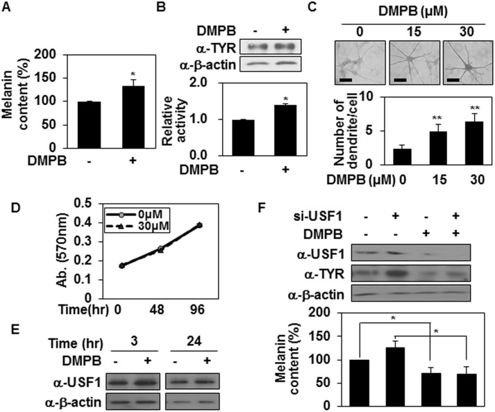
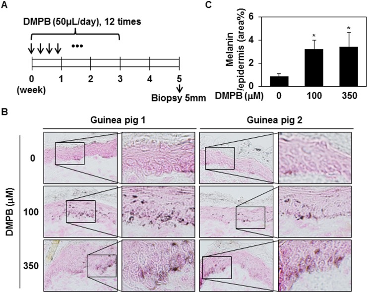
We investigated the potential melanogenic effect of compounds from Zingiber cassumunar Roxb. Our data revealed that chloroform-soluble extract of Z. cassumunar enhanced melanin synthesis in B16F10 melanoma cells. Among the components of the chloroform extract, (E)-4-(3,4-dimethoxyphenyl)but-3-en-1-ol (DMPB) increased melanogenesis in both B16F10 cells and human primary melanocytes. In B16F10 cells, DMPB enhanced the activation of ERK and p38, and the level of tyrosinase. Although the level of microphthalmia-associated transcription factor was unchanged in DMPB-treated B16F10 cells, DMPB increased levels and nuclear localization of upstream stimulating factor-1 (USF1). Consistently, DMPB-mediated melanin synthesis was diminished in USF1-knockdown cells. Furthermore, DMPB induced hyperpigmentation in brown guinea pigs in vivo. Together, these data suggest that DMPB may promote melanin synthesis via USF1 dependent fashion and could be used as a clinical therapeutic agent against hypopigmentation-associated diseases.


Conflict of interest statement

Competing Interests: The authors have declared that no competing interests exist.

Figures

Fig 1
Fig 1. (E)-4-(3,4-Dimethoxyphenyl)but-3-en-1-ol from Z. cassumunar enhances melanin synthesis.
(A) The methanol extract of Z. cassumunar was partitioned with hexanes, chloroform, and butanol (top panel). B16F10 cells were treated with three fractions of Z. cassumunar (BF: Butanol fraction, CF: Chloroform fraction, HF: Hexane fraction; 20 μg/ml, 48hr). The melanin contents were analyzed by measuring the absorbance at 405 nm (bottom panel). DMSO was used as a control. The mean percentages of melanin content are shown. **, p < 0.01 versus DMSO treated cells. (B) B16F10 cells were treated with the indicated compounds extracted from Z. cassumunar (30 μM each) for 48 hr, and the melanin contents were determined. *, p < 0.05 versus DMSO treated cells. (C,D) B16F10 cells were treated with either various concentrations of DMPB for 48 hr (C) or with 30 μM of DMPB for the indicated times (D), and the mean percentages of melanin content are shown. (E) B16F10 cells were treated with of 30 μM of DMPB or 1 μM of α-MSH for 48 hr. The mean percentages of melanin content are shown. **, p < 0.01 versus DMSO treated cells.
Fig 2
Fig 2. DMPB increases tyrosinase expression but not tyrosinase activity.
(A) B16F10 cells were treated with 30 μM of DMPB for 48 hr, and mRNA level of tyrosinase was analyzed by RT-PCR (top panel). Total cell lysate was extracted and tyrosinase levels were measured by Western blot analysis. The relative density of tyrosinase(TYR) was quantitated using Image Studio software (middle panel). The mean percentages of tyrosinase density ± SD are shown *, p < 0.05 versus DMSO treated cells. DMPB-treated B16F10 cells (30 μM, 48 hr) were lysed. Cell lysates (100 μg) were reacted with L-DOPA at 37°C for 2 hr, and tyrosinase activity was determined at 470 nm (bottom panel). The mean percentages of tyrosinase activity ± SD are shown **, p < 0.01 versus DMSO treated cells. (B) DMPB-treated B16F10 cells (48 hr) were reacted with L-DOPA at 37°C for 30 min. Bright-field microscopic images are shown. Scale bars = 50 μm. (C) Cell lysates (20 μg and 40 μg) from B16F10 cells treated with the indicated concentrations of DMPB were subjected to Western blot analysis using an anti-tyrosinase antibody (top panel) or reacted with L-DOPA at 37°C for 2 hr to determine tyrosinase activity (bottom panel). The mean percentages of tyrosinase activity ± SD are shown. (D) B16F10 cells were incubated with various concentrations of DMPB for the indicated time periods, and cell viability was determined by MTT assay. Percentage values were compared between treated and untreated (control). Data are expressed as mean ± SD for three independent experiments.
Fig 3
Fig 3. MAP kinases are involved in DMPB-mediated melanogenic control.
(A) B16F10 cells were treated with 30 μM of DMPB for the indicated time periods, and the phosphorylation of p38 and ERK and levels of tyrosinase were analyzed by Western blot analysis. (B) B16F10 cells were treated with the indicated amounts of DMPB for 48 hr, and the phosphorylation of p38 and ERK and levels of tyrosinase were analyzed by Western blot analysis. (C) B16F10 cells were preincubated with (+) or without (-) the inhibitor (1 μM of PD98059, 10 μM of U0126) for 1 hr, then treated with 30 μM of DMPB for 48 hr, and Western blot analysis was performed with the indicated antibodies (top panel). The melanin contents were analyzed by measuring the absorbance at 405 nm (bottom panel). The mean percentages of melanin content are shown *, p < 0.05 versus DMSO treated cells. (D) B16F10 cells were preincubated with (+) or without (-) p38 inhibitors (5 μM of SB239063 for 30 min, 10 μM of SB203580 for 1 hr) and treated with 30 μM of DMPB for 48 hr, and Western blot analysis was performed with the indicated antibodies (top panel). The melanin contents were analyzed by measuring the absorbance at 405 nm (bottom panel). The mean percentages of melanin content are shown *, p < 0.05 versus DMSO treated cells.
Fig 4
Fig 4. DMPB stimulates melanin synthesis by activating USF1.
(A) B16F10 cells were treated with 30 μM of DMPB for the indicated time periods. Cytoplasmic and nuclear fractions were isolated and analyzed by Western blotting. (B) B16F10 cells were transfected with si-CON or si-MITF for 48 hr. MITF protein levels were analyzed by Western blotting in the absence or presence of 1 μM α-MSH for 3 hr. (C) Tyrosinase protein levels were analyzed by Western blotting in the absence or presence of 30 μM DMPB for 48 hr (top panel) and the melanin content was measured by absorbance at 405 nm (bottom panel). The mean percentages of melanin content are shown. **, p < 0.01 versus DMSO treated cells. (D) B16F10 cells were treated with 30 μM of DMPB for the indicated time periods in the presence of 1 μM α-MSH. MITF and p53 protein levels were analyzed by Western blotting and MITF mRNA level was analyzed by RT-PCR. (E) B16F10 cells were treated with DMPB for 3 hr, and Immunofluorescence analysis was performed with anti-USF1 antibody. Scale bar = 20 μm (F) B16F10 cells were transfected with si-con or si-USF1 and treated with DMPB (30 μM) for 48 hr. Indicated protein levels were analyzed by Western blotting (top panel) and the melanin content was measured by absorbance at 405 nm (bottom panel).**, p<0.01; *, p < 0.05 versus DMSO treated cells.
Fig 5
Fig 5. DMPB promotes melanin synthesis in human melanocytes.
(A, B) Human melanocytes were treated with 30 μM of DMPB. After 48 hr melanin contents were analyzed by measuring the absorbance at 405 nm (A). The mean percentages of melanin content are shown *, p < 0.05 versus DMSO treated cells. Tyrosinase expression levels were analyzed by Western blot analysis (B; top panel). Cell lysates (100 μg) were reacted with L-DOPA at 37°C for 2 hr, and tyrosinase activity was determined at 470 nm (B; bottom panel). The mean percentages of tyrosinase activity ± SD are shown. *, p < 0.05 versus DMSO treated cells. (C) Human melanocytes were plated on 12-well plates, treated with the indicated concentrations of DMPB for 48 hr, and reacted with L-DOPA at 37°C for 30 min. Bright-field microscopic images are shown (top panel). Scale bars = 50 μm. The mean percentages of the number of dendrite/cell ± SD are shown (bottom panel). **, p < 0.01 versus DMSO treated cells. (D) Primary melanocytes were incubated with DMPB (30 μM) in a 96-well plate for the indicated periods, and cell viability was determined by MTT-based spectrophotometric assay. Percentage values were compared between treated and untreated (control) cells. The data are expressed as mean ± SD from three independent experiments. (E) Primary melanocytes were treated with 30 μM of DMPB for the indicated periods. USF1 protein levels were analyzed by Western blotting. (F) Primary melanocytes were transfected with USF1 targeting siRNA and treated with DMPB (30 μM). After 48 hr, tyrosinase and USF1 expression levels were analyzed by Western blot analysis (top panel). Cells were harvested and melanin contents were analyzed by measuring the absorbance at 405 nm (bottom panel). The mean percentages or melanin content are shown *, p < 0.05.
Fig 6
Fig 6. DMPB enhances hyperpigmentation in brown guinea pigs.
(A) The dorsal skins of guinea pigs were topically treated with DMSO (control), 100 μM DMPB, or 350 μM DMPB (50 μl each) 12 times in 3 weeks. On day 35 (after the first treatment), skin specimens were obtained by a 5-mm punch biopsy. (B) Frozen skin specimens were cut at 10- μM thickness, and melanin pigment was visualized by Fontana-Masson staining. Original magnification, X200. (C) Bar graph showing the mean percentage of melanin ± SD in the epidermis. Fontana-Masson-stained melanin in three randomly selected fields was measured with the ImageJ program. *, p < 0.05 versus DMSO treated skins.

References

    1. Yamaguchi Y, Takahashi K, Zmudzka BZ, Kornhauser A, Miller SA, Tadokoro T, et al. Human skin responses to UV radiation: pigment in the upper epidermis protects against DNA damage in the lower epidermis and facilitates apoptosis. FASEB J. 2006; 20: 1486–1488. - PubMed
    1. Slominski A, Tobin DJ, Shibahara S, Wortsman J. Melanin pigmentation in mammalian skin and its hormonal regulation. Physiol. Rev. 2004; 84: 1155–1228. - PubMed
    1. Cheli Y, Ohanna M, Ballotti R, Bertolotto C. Fifteen-year quest for microphthalmia-associated transcription factor target genes. Pigment Cell Melanoma Res. 2010; 23: 27–40. 10.1111/j.1755-148X.2009.00653.x - DOI - PubMed
    1. Galibert MD, Carreira S, Goding CR. The Usf-1 transcription factor is a novel target for the stress-responsive p38 kinase and mediates UV-induced Tyrosinase expression. EMBO J. 2001; 20: 5022–5031. - PMC - PubMed
    1. Aksan I, Goding CR. Targeting the microphthalmia basic helix-loop-helix-leucine zipper transcription factor to a subset of E-Box elements in vitro and in vivo. Mol. Cell Biol. 1998; 18: 6930–6938. - PMC - PubMed

Publication types

MeSH terms