. 2015 Nov 4;10(11):e0141783.
doi: 10.1371/journal.pone.0141783. eCollection 2015.

Ocular Hypotensive Effects of the ATP-Sensitive Potassium Channel Opener Cromakalim in Human and Murine Experimental Model Systems

Affiliations

Ocular Hypotensive Effects of the ATP-Sensitive Potassium Channel Opener Cromakalim in Human and Murine Experimental Model Systems

Uttio Roy Chowdhury et al. PLoS One. .

Erratum in

Abstract

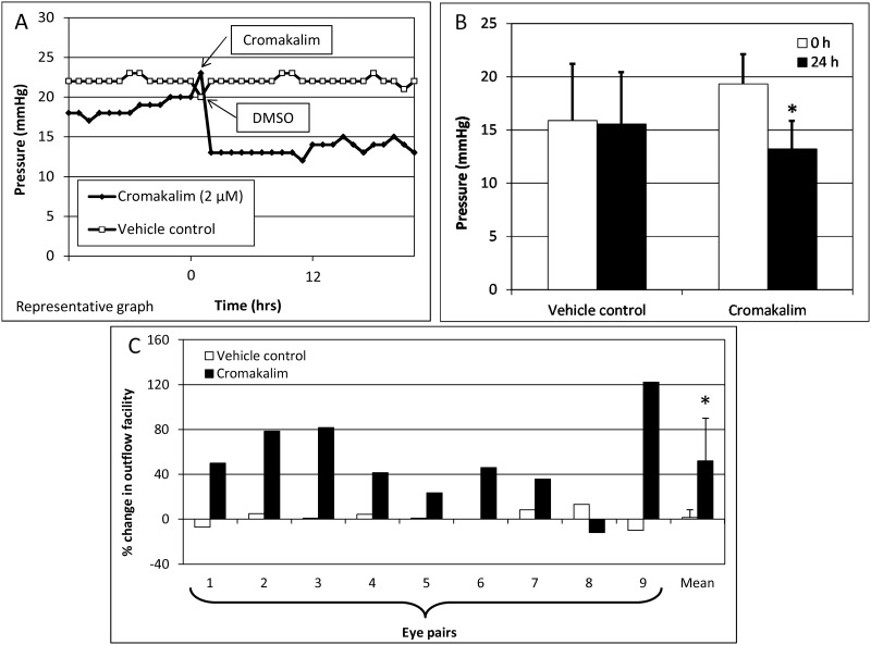
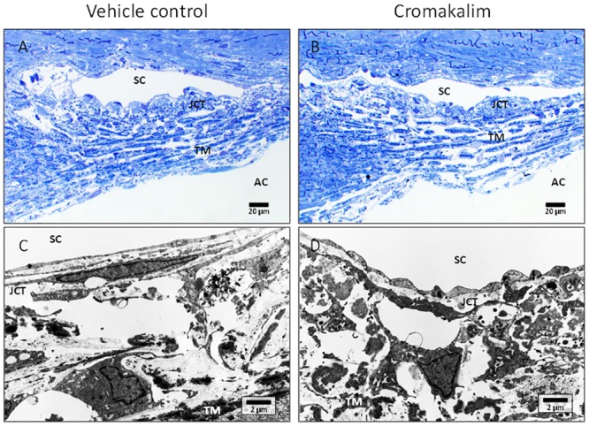
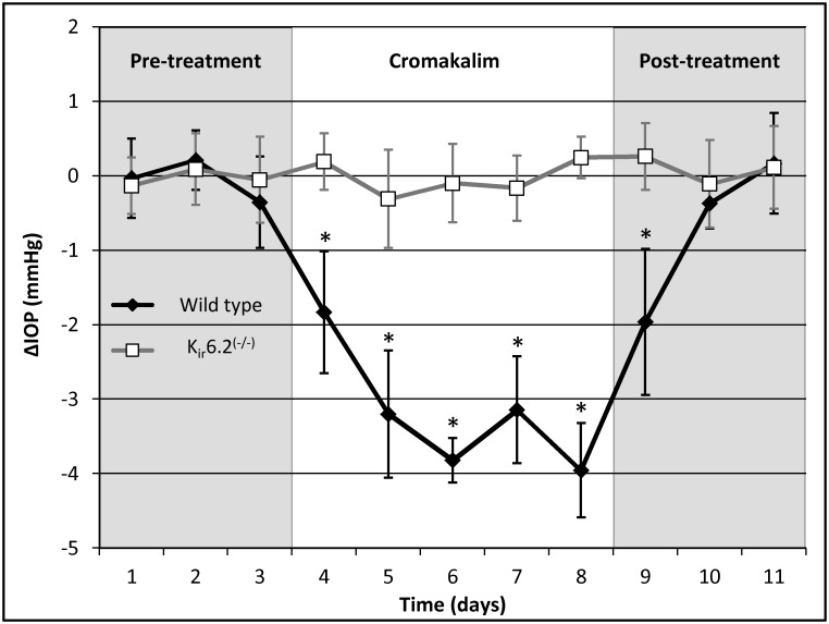
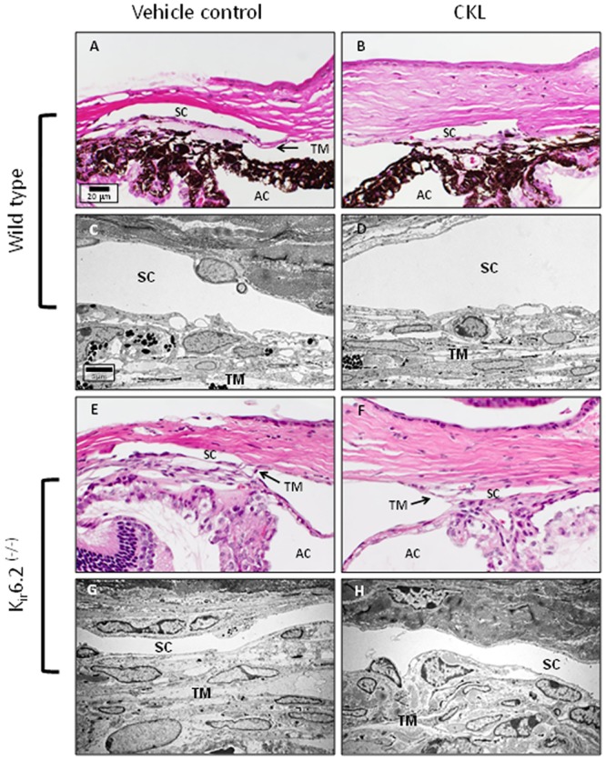
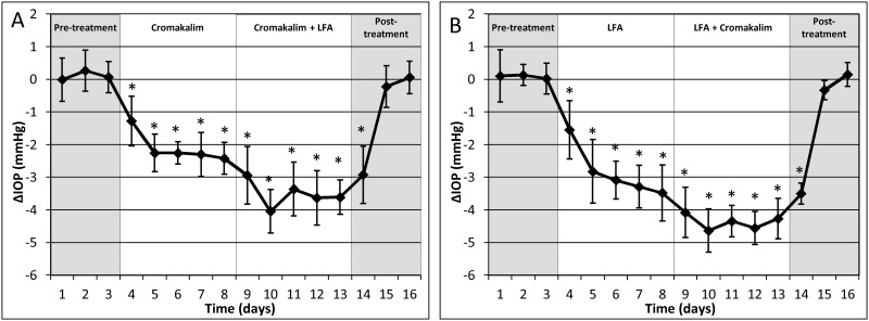
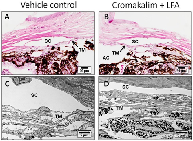
Elevated intraocular pressure (IOP) is the most prevalent and only treatable risk factor for glaucoma, a leading cause of irreversible blindness worldwide. Unfortunately, all current therapeutics used to treat elevated IOP and glaucoma have significant and sometimes irreversible side effects necessitating the development of novel compounds. We evaluated the IOP lowering ability of the broad spectrum KATP channel opener cromakalim. Cultured human anterior segments when treated with 2 μM cromakalim showed a decrease in pressure (19.33 ± 2.78 mmHg at 0 hours to 13.22 ± 2.64 mmHg at 24 hours; p<0.001) when compared to vehicle treated controls (15.89 ± 5.33 mmHg at 0 h to 15.56 ± 4.88 mmHg at 24 hours; p = 0.89). In wild-type C57BL/6 mice, cromakalim reduced IOP by 18.75 ± 2.22% compared to vehicle treated contralateral eyes (17.01 ± 0.32 mmHg at 0 hours to 13.82 ± 0.37 mmHg at 24 hours; n = 10, p = 0.002). Cromakalim demonstrated an additive effect when used in conjunction with latanoprost free acid, a common ocular hypotensive drug prescribed to patients with elevated IOP. To examine KATP channel subunit specificity, Kir6.2(-/-) mice were treated with cromakalim, but unlike wild-type animals, no change in IOP was noted. Histologic analysis of treated and control eyes in cultured human anterior segments and in mice showed similar cell numbers and extracellular matrix integrity within the trabecular meshwork, with no disruptions in the inner and outer walls of Schlemm's canal. Together, these studies suggest that cromakalim is a potent ocular hypotensive agent that lowers IOP via activation of Kir6.2 containing KATP channels, its effect is additive when used in combination with the commonly used glaucoma drug latanoprost, and is not toxic to cells and tissues of the aqueous humor outflow pathway, making it a candidate for future therapeutic development.


Conflict of interest statement

Competing Interests: The authors have declared that no competing interests exist.

Figures

Fig 1
Fig 1. The KATP channel opener cromakalim has ocular hypotensive activity in perfusion cultures of human anterior segments.
A) Representative graph shows reduction in pressure following addition of cromakalim (2 μM). B) Treatment with 2 μM cromakalim caused significant decrease in pressure when compared to vehicle (DMSO) treated controls (n = 9). C) Outflow facility in all eye pairs treated with cromakalim for 24 hours along with the mean value for the group. Outflow facility was increased in 8 out of the 9 eye pairs. Data represents mean ± standard deviation. *p<0.001.
Fig 2
Fig 2. Histologic analysis of human anterior segment ocular tissue following treatment with cromakalim.
A, B) 1 μm toluidine blue stained representative sections of vehicle (DMSO) and cromakalim treated eyes. C, D) Transmission electron micrographs showing ultrastructure of vehicle and cromakalim treated eyes. Vehicle and treated eyes had similar morphology and ultrastructural appearance showing no detrimental side effects of cromakalim treatment. SC = Schlemm’s canal; TM = trabecular meshwork; AC = anterior chamber; JCT = juxtacanalicular region.
Fig 3
Fig 3. Cromakalim lowered intraocular pressure (IOP) in wild type C57BL/6 mice but not in Kir6.2(-/-) mice.
IOP was significantly reduced within 24 hours of 5 mM cromakalim treatment. Average IOP reduction was calculated to be 3.19 ± 0.41 mmHg over a treatment period of 5 days in wild type mice (n = 10). IOP was found to return to baseline levels within 48 hours of termination of treatment. In contrast, Kir6.2(-/-) mice (n = 10) showed no change in IOP following cromakalim treatment. IOP in these animals remained at baseline levels even after topical administration of cromakalim. Values correspond to average difference (in mmHg) between treated and control eyes for individual days (ΔIOP). *p<0.01
Fig 4
Fig 4. Representative images of the conventional outflow pathway of C57BL/6 (A-D) and Kir6.2(-/-) (E-H) treated and vehicle control mice.
Similar to human anterior segments, comparison between vehicle and cromakalim treated eyes showed no observable cell or tissue changes. Both vehicle and cromakalim treated groups showed normal morphology and ultrastructure with intact inner and outer walls of Schlemm’s canal, viable cells and an evenly distributed extracellular matrix. SC = Schlemm’s canal; TM = trabecular meshwork; AC = anterior chamber.
Fig 5
Fig 5. Combination treatment with cromakalim and latanoprost free acid (LFA) showed an additive effect in intraocular pressure (IOP) reduction, irrespective of which drug was used to treat the eyes first.
A) Treatment with cromakalim (5 mM) + LFA (0.1 mM) showed an additional IOP reduction of 78.52 ± 47.57% (n = 10, p = 0.002) when compared to treatment with cromakalim alone. B) Combination of cromakalim + LFA showed an additional IOP reduction of 55.74 ± 19.32% (n = 10, p = 0.002) compared to treatment with LFA alone. Values correspond to average difference (in mmHg) between treated and control eyes for individual days (ΔIOP). *p<0.01.
Fig 6
Fig 6. Representative transmission electron micrographs of mouse eyes treated with cromakalim + latanoprost free acid (LFA) or vehicle.
Comparison of the micrographs show normal anatomy and ultrastructure in vehicle (A, C) and cromakalim + LFA treated tissue (B, D), indicating no observable changes due to treatment. SC = Schlemm’s canal; TM = trabecular meshwork; AC = anterior chamber.

References

    1. Quigley HA, Broman AT. The number of people with glaucoma worldwide in 2010 and 2020. Br J Ophthalmol. 2006;90(3):262–7. . - PMC - PubMed
    1. Weinreb RN, Aung T, Medeiros FA. The pathophysiology and treatment of glaucoma: a review. JAMA: the journal of the American Medical Association. 2014;311(18):1901–11. Epub 2014/05/16. 10.1001/jama.2014.3192 . - DOI - PMC - PubMed
    1. Tham YC, Li X, Wong TY, Quigley HA, Aung T, Cheng CY. Global Prevalence of Glaucoma and Projections of Glaucoma Burden through 2040: A Systematic Review and Meta-Analysis. Ophthalmology. 2014. Epub 2014/07/01. 10.1016/j.ophtha.2014.05.013 . - DOI - PubMed
    1. Friedman DS, Wolfs RC, O'Colmain BJ, Klein BE, Taylor HR, West S, et al. Prevalence of open-angle glaucoma among adults in the United States. Arch Ophthalmol. 2004;122(4):532–8. Epub 2004/04/14. 10.1001/archopht.122.4.532 - DOI - PMC - PubMed
    1. Weinreb RN, Khaw PT. Primary open-angle glaucoma. Lancet. 2004;363(9422):1711–20. Epub 2004/05/26. 10.1016/S0140-6736(04)16257-0 . - DOI - PubMed

Publication types

MeSH terms