. 2015 Nov 4;10(11):e0142093.
doi: 10.1371/journal.pone.0142093. eCollection 2015.

Loss of Metabotropic Glutamate Receptor 5 Function on Peripheral Benzodiazepine Receptor in Mice Prenatally Exposed to LPS

Affiliations

Loss of Metabotropic Glutamate Receptor 5 Function on Peripheral Benzodiazepine Receptor in Mice Prenatally Exposed to LPS

Dany Arsenault et al. PLoS One. .

Abstract

Parental microglial induced neuroinflammation, triggered by bacterial- or viral infections, can induce neuropsychiatric disorders like schizophrenia and autism to offspring in animal models. Recent investigations suggest that microglia, the resident immune cells of the brain, provides a link between neurotransmission, immune cell activation, brain inflammation and neuronal dysfunction seen with the offspring. Relatively little is known about how reduction of brain inflammation and restoration of glial function are associated with diminution of brain degeneration and behavioral deficits in offspring. Increased mGluR5 expression and the long-lasting excitotoxic effects of the neurotoxin during brain development are associated with the glial dysfunctions. We investigated the relationship of mGluR5 and PBR and how they regulate glial function and inflammatory processes in mice prenatally exposed to LPS (120μg/kg, between gestational days 15 and 17), an inflammatory model of a psychiatric disorder. Using PET imaging, we showed that pharmacological activation of mGluR5 during 5 weeks reduced expression of classic inflammation marker PBR in many brain areas and that this molecular association was not present in LPS-exposed offspring. The post-mortem analysis revealed that the down regulation of PBR was mediated through activation of mGluR5 in astrocytes. In addition, we demonstrated that this interaction is defective in a mouse model of the psychiatric deficit offering a novel insight of mGluR5 involvement to brain related disorders and PBR related imaging studies. In conclusion, mGluR5 driven glutamatergic activity regulates astrocytic functions associated with PBR (cholesterol transport, neurosteroidogenesis, glial phenotype) during maturation and could be associated with neuropsychiatric disorders in offspring.


Conflict of interest statement

Competing Interests: The authors have declared that no competing interests exist.

Figures

Fig 1
Fig 1. Time lines of the experiments.
Pregnant mice received an injection of 120 μg/kg of LPS or an equivalent volume of saline between GD15 to GD17. Expression of mGluR5 (PnD37-PnD39) and PBR (PnD42-PnD44) in the brain of the offspring prenatally exposed to LPS or saline were evaluated by PET imaging. Animals of both groups (LPS or saline) were then exposed to treatment during 5 weeks (from PnD56 to PnD91). Three types of treatments were tested: CDPPB (10 mg/kg; mGluR5 agonist), MTEP (3 mg/kg; mGluR5 antagonist) and control (solvent alone). Finally, the in vivo expression of mGluR5 (PnD117-119) and PBR (PnD125-127) were re-evaluated after the treatments and the animals were sacrificed a few days after the last imaging session (PnD128-130) for post-mortem analysis. Abbreviations: GD, gestational day; [11C]PBR28, peripheral benzodiazepine receptor 28; [18F]FPEB, [18F]fluoro-5-(2-pyridinylethynyl)benzonitrile; PnD, postnatal day.>>
Fig 2
Fig 2. Effect of aging and different prenatal treatments on mGluR5 expression investigated with PET imaging using [18F]FPEB.
PET imaging revealed a significant increase of [18F]FPEB binding during maturation (from PnD37-39 to PnD 117–119) in the striatum and the whole brain of the mice prenatally exposed to saline (A), while in the LPS-exposed offspring the [18F]FPEB binding increased in the cortex, hippocampus, striatum and the whole brain during the same maturation period (B). During the adolescence (PnD37-39) we observed a lower binding of [18F]FPEB in the striatum and the whole brain of the LPS-exposed offspring compared to the saline-treated offspring (C). However, [18F]FPEB binding was similar in both groups at PnD117-119 (D). (E) Coronal slices of hippocampal and striatal level of [18F]FPEB in offspring prenatally exposed to saline or LPS at PnD 37–39 and PnD 117–119. Values are expressed as mean ± SEM. Abbreviations: [18F]fluoro-5-(2-pyridinylethynyl)benzonitrile; Ctx, cortex; Hip, hippocampus; Hth, hypothalamus; OB, olfactory bulb; PnD, postnatal day; Str, striatum; W, whole brain. *p < 0.05, **p < 0.01 >>
Fig 3
Fig 3. Effects of postnatal CDPPB treatment on [18F]FPEB binding potential.
CDPPB treatment did not change the [18F]FPEB binding potential in the quantified brain region of the offspring prenatally exposed to saline solution (A). However, the LPS-exposed offspring had a lower [18F]FPEB accumulation in the cortex and hippocampus following CDPPB treatment (B). (C) Axial view of a representative mouse from each group. Values are expressed as mean ± SEM. Abbreviations: CDPPB, 3-cyano-N-(1,3-diphenyl-1H-pyrazol-5-yl)benzamide; [18F]FPEB, [18F]fluoro-5-(2-pyridinylethynyl)benzonitrile; Ctx, cortex; Hip, hippocampus; Hth, hypothalamus; OB, olfactory bulb; PnD, postnatal day; Str, striatum; W, whole brain. *p < 0.05, **p < 0.01. Statistical analyses were performed using one-sample t test (all comparisons of panel A; OB, Hip, Str, Hth and W of panel B) or Wilcoxon signed-rank test (Ctx of panel B). The number of animals was 10–11 for the saline group and 12 for the LPS-exposed group. >>
Fig 4
Fig 4. Postnatal CDPPB treatment decreased [11C]PBR accumulation in saline-exposed offspring, but not in offspring prenatally exposed to LPS.
Postnatal CDPPB treatment decreased the accumulation of [11C]PBR in the olfactory bulb, cerebellum, hippocampus, striatum, hypothalamus and the whole brain of the offspring prenatally exposed to saline solution (A). However, [11C]PBR accumulation did not change in any quantified brain region of the LPS-exposed offspring (B). Axial PET images of [11C]PBR accumulation at the midbrain level illustrate the decreased accumulation after CDPPB treatment in the prenatally saline-exposed offspring while there is no significant change in the brain of CDPPB treated mice prenatally exposed to LPS (C). Values are expressed as mean ± SEM. Abbreviations: CDPPB, 3-cyano-N-(1,3-diphenyl-1H-pyrazol-5-yl)benzamide; [11C] PBR28, peripheral benzodiazepine receptor 28; Ctx, cortex; Crbl, cerebellum; Hip, hippocampus; Hth, hypothalamus; OB, olfactory bulb; PnD, postnatal day; Str, striatum; W, whole brain. *p < 0.05, **p < 0.01 >>
Fig 5
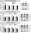
Fig 5. Astrocytic marker was decreased in the brain of prenatally saline-exposed offspring, but not in the offspring prenatally exposed to LPS, following pharmacological activation of mGluR5.
A prenatal administration of LPS did not change the brain expression level of iba1, CD68, GFAP or mGluR5 in 4-month-old mice (A). Postnatal CDPPB treatment reduced the GFAP level, without effect on iba1, CD68 or mGluR5 in the mice prenatally exposed to saline solution. The quantified molecular markers did not change in the brain of mice treated with MTEP (B). No change in iba1, CD68, GFAP or mGluR5 was observed in the brain of the mice prenatally exposed to LPS (C). Values are expressed as mean ± SEM. Abbreviations: MTEP, 3-((2-methyl-4-thiazolyl)ethynyl)pyridine; CDPPB, 3-cyano-N-(1,3-diphenyl-1H-pyrazol-5-yl) benzamide; CD68, cluster of differentiation 68); iba1, ionized calcium binding adaptor molecule-1; GFAP, glial fibrillary acidic protein; mGluR5, metabotropic glutamate receptor subtype 5; ROD, relative optical density. **p < 0.01 >>

References

    1. Niswender CM, Conn PJ (2010) Metabotropic glutamate receptors: physiology, pharmacology, and disease. Annu Rev Pharmacol Toxicol 50: 295–322. 10.1146/annurev.pharmtox.011008.145533 - DOI - PMC - PubMed
    1. Hermans E, Challiss RA (2001) Structural, signalling and regulatory properties of the group I metabotropic glutamate receptors: prototypic family C G-protein-coupled receptors. Biochem J 359: 465–484. - PMC - PubMed
    1. Hovelso N, Sotty F, Montezinho LP, Pinheiro PS, Herrik KF, Mork A (2012) Therapeutic potential of metabotropic glutamate receptor modulators. Curr Neuropharmacol 10: 12–48. 10.2174/157015912799362805 - DOI - PMC - PubMed
    1. Pellegrino D, Cicchetti F, Wang X, Zhu A, Yu M, Saint-Pierre M, et al. (2007) Modulation of dopaminergic and glutamatergic brain function: PET studies on parkinsonian rats. J Nucl Med 48: 1147–1153. - PubMed
    1. Ayala JE, Chen Y, Banko JL, Sheffler DJ, Williams R, Telk AN, et al. (2009) mGluR5 positive allosteric modulators facilitate both hippocampal LTP and LTD and enhance spatial learning. Neuropsychopharmacology 34: 2057–2071. 10.1038/npp.2009.30 - DOI - PMC - PubMed

Publication types

MeSH terms