. 2015 Nov 12;527(7577):179-85.
doi: 10.1038/nature15698. Epub 2015 Nov 4.

Basomedial amygdala mediates top-down control of anxiety and fear

Affiliations

Basomedial amygdala mediates top-down control of anxiety and fear

Avishek Adhikari et al. Nature. .

Abstract

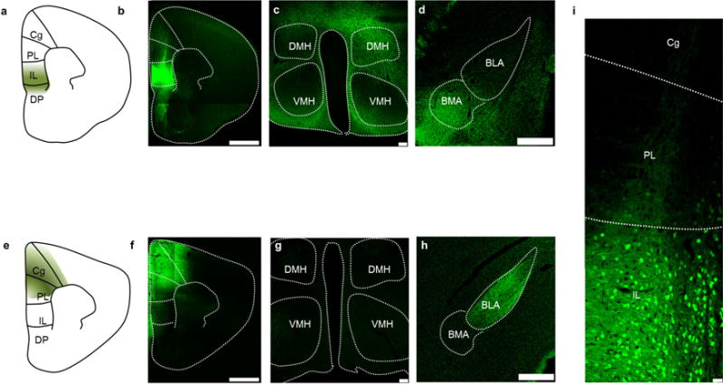
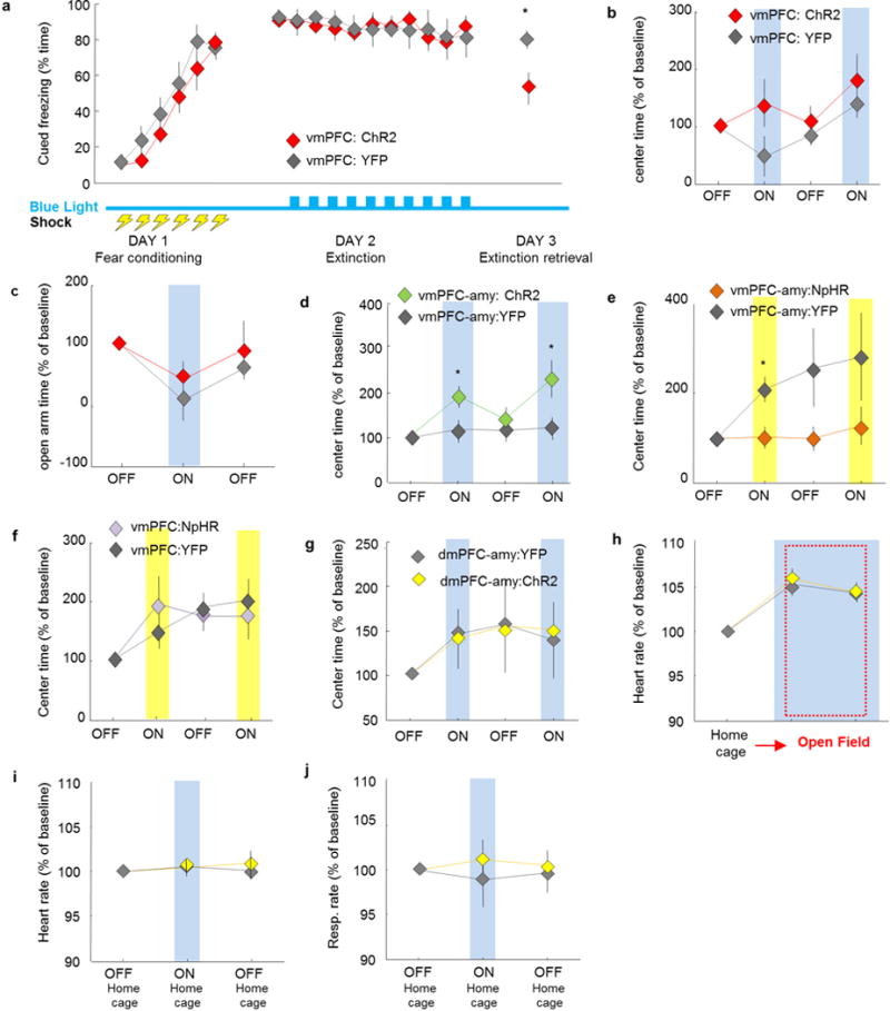
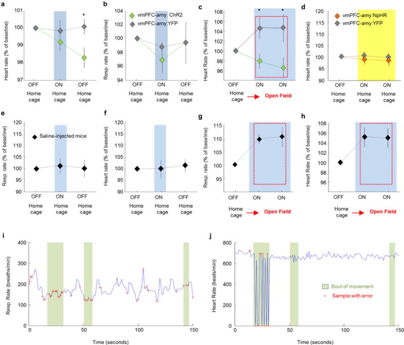
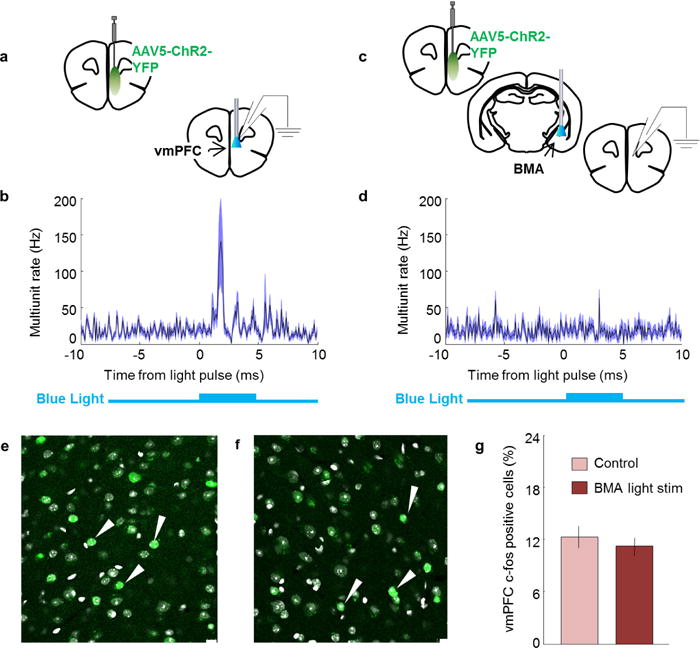
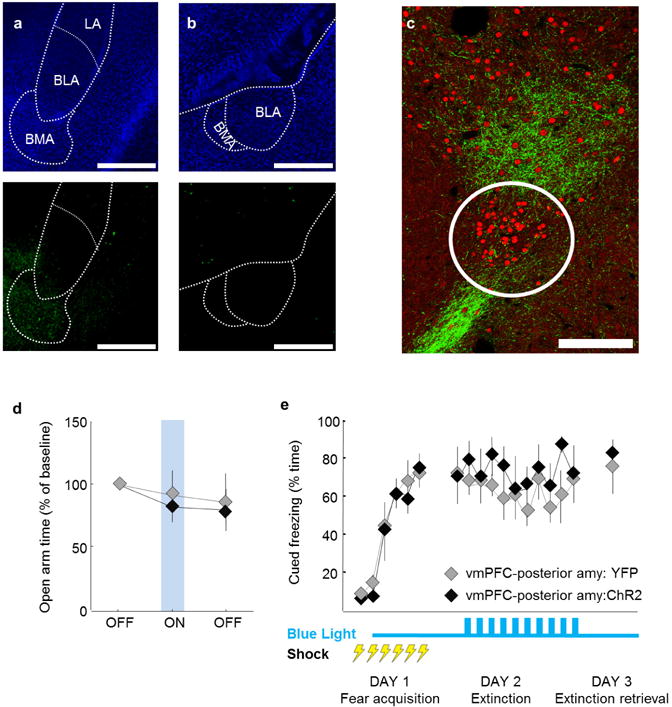
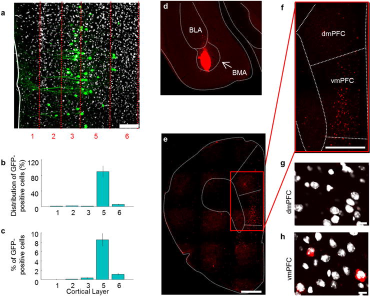
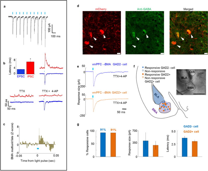
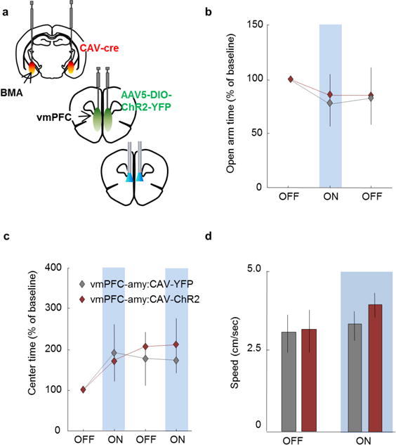
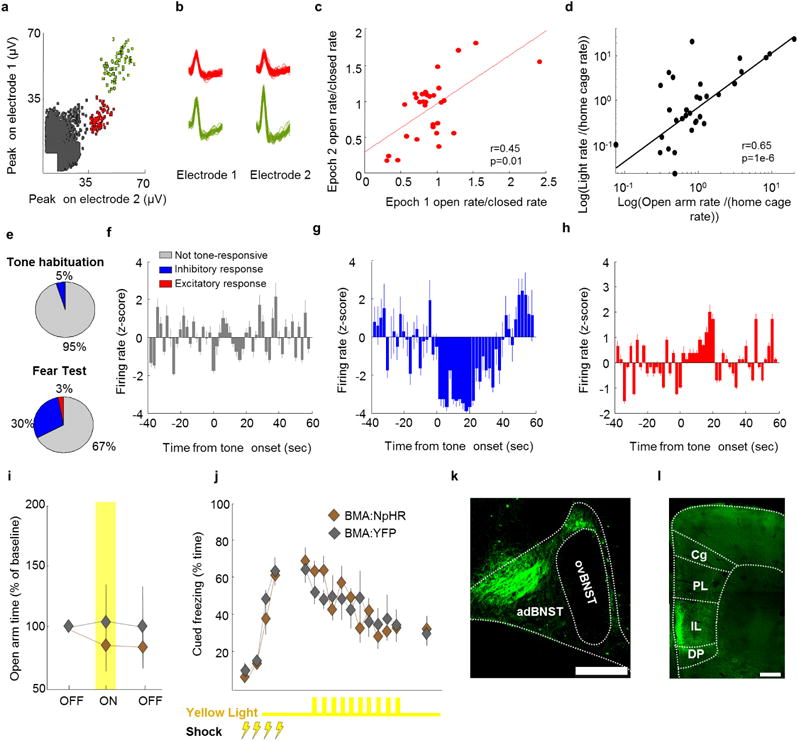
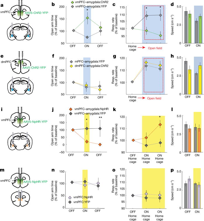
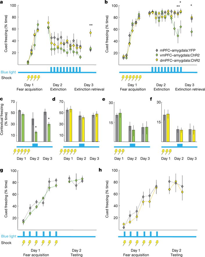
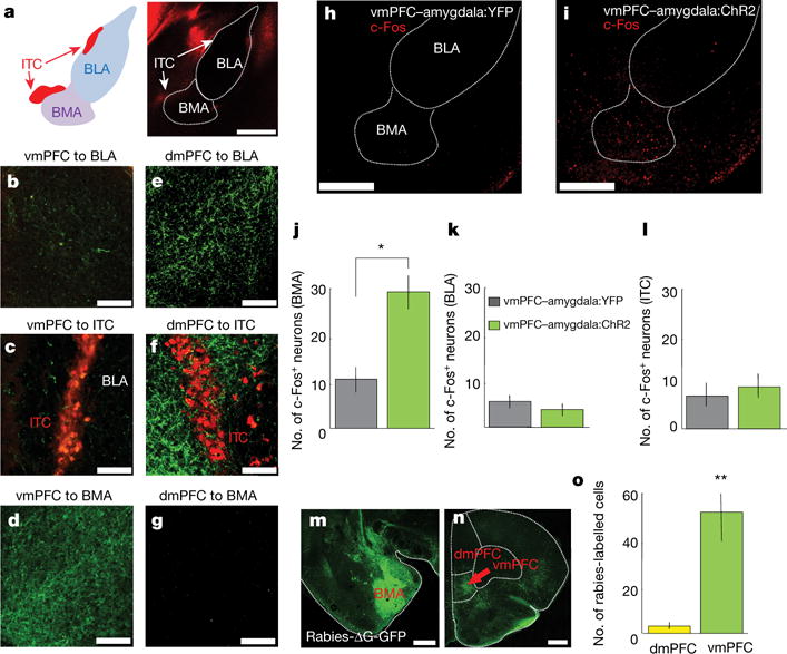
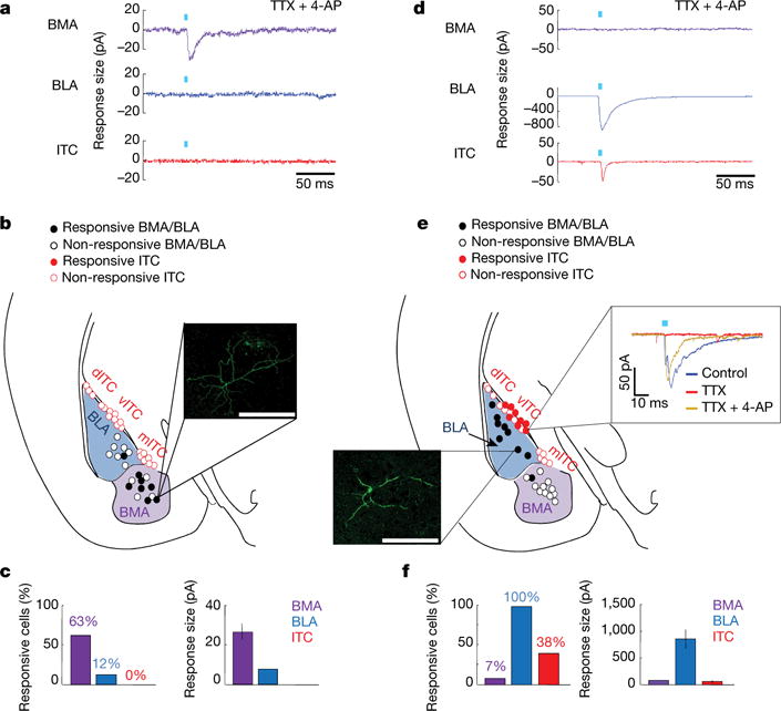
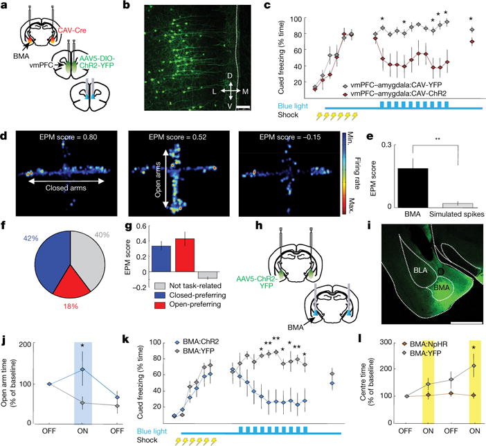
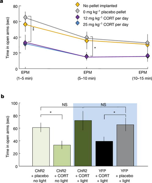
Anxiety-related conditions are among the most difficult neuropsychiatric diseases to treat pharmacologically, but respond to cognitive therapies. There has therefore been interest in identifying relevant top-down pathways from cognitive control regions in medial prefrontal cortex (mPFC). Identification of such pathways could contribute to our understanding of the cognitive regulation of affect, and provide pathways for intervention. Previous studies have suggested that dorsal and ventral mPFC subregions exert opposing effects on fear, as do subregions of other structures. However, precise causal targets for top-down connections among these diverse possibilities have not been established. Here we show that the basomedial amygdala (BMA) represents the major target of ventral mPFC in amygdala in mice. Moreover, BMA neurons differentiate safe and aversive environments, and BMA activation decreases fear-related freezing and high-anxiety states. Lastly, we show that the ventral mPFC-BMA projection implements top-down control of anxiety state and learned freezing, both at baseline and in stress-induced anxiety, defining a broadly relevant new top-down behavioural regulation pathway.


Conflict of interest statement

The authors declare no competing financial interests.

Figures

Extended Data Figure 1
Extended Data Figure 1. Methodology necessary for targeting viral infusions restricted to vmPFC and dmPFC
a, Scheme showing subregions of the mPFC. b, Representative example from one mouse showing a coronal section containing the vmPFC (IL+ DP) in a mouse expressing YFP in the vmPFC. c, d, Coronal section depicting vmPFC fibres in the hypothalamus (c) and amygdala (d). n = example from 1 mouse chosen from n = 7 mice (a–d). e–h, Same as a–d, but for an animal that received viral injection in the dmPFC (PL and Cg). n = example from 1 mouse chosen from n = 7 mice (e–h). c, g, Note the presence of fibres from the vmPFC, but not dmPFC, in the hypothalamus. Specifically, the vmPFC projects strongly to the dorsomedial, but not ventromedial hypothalamus (DMH and VMH, respectively). g, Section showing expression of YFP in a vmPFC:YFP mouse. n = example from 1 mouse chosen from n = 7 mice. Cg, cingulate cortex; PL, prelimbic cortex; IL, infralimbic cortex; D P, dorsal peduncular cortex. Scale bars, 1 mm (b, f); 100 μm (c, g); 500 μm (d, h); 35 μm (i).
Extended Data Figure 2
Extended Data Figure 2. Optogenetic manipulations involving the mPFC and its projections to the amygdala in fear and anxiety paradigms
a, Mice were fear conditioned to six tone-shock (0.7 mA, 2-s shocks) pairings on day 1 (fear acquisition). On day 2 (fear extinction) animals were exposed to the tone in a different context for 11 trials. Blue light was delivered only on day 2, for trials 2–11. vmPFC:ChR2 mice froze less than control mice during day 3 (extinction retrieval). b, c, Blue light delivery did not change avoidance of open spaces in vmPFC:ChR2 mice relative to control mice in the open field (b) or the EPM (c). n = 7 vmPFC:ChR2 and 7 vmPFC:YFP mice (a–c). d, Excitation of the vmPFC–amygdala projection with blue light increased exploration of the centre of the open field in vmPFC–amygdala:ChR2 mice relative to controls. Two-way repeated measures ANOVA, opsin × epoch interaction, F3,68 = 3.1, P = 0.03, post hoc Wilcoxon rank sum test P = 0.002. n = 12 vmPFC–amygdala:ChR2 and 13 vmPFC–amygdala:YFP mice. e, Inhibition of the same projection in vmPFC–amygdala:NpHR mice with yellow light increased avoidance of open spaces. Two-way repeated-measures ANOVA, main effect of opsin, F3,68 = 7.26, P = 0.008, post-hoc Wilcoxon rank sum test P = 0.03. n = 14 vmPFC–amygdala:NpHR and 11 vmPFC–amygdalal:YFP mice. f, Same as e, but for inhibition of vmPFC cell bodies. n = 11 vmPFC:NpHR and 8 vmPFC:YFP mice. g–j, Optogenetic stimulation of the dmPFC–amygdala projection did not alter behaviour in the open field (g), heart rate in the open field (h), heart rate in the home cage (i), or respiration rates in the home cage (j). Blue light stimulation epochs are labelled ON and/or with a blue bar. n = 7 dmPFC–amygdala:YFP and 7 mPFC–amygdala:ChR2 mice. b–j, Data are plotted in 5-min consecutive intervals. Light delivery epochs are labelled ON and/or with a blue or yellow bar. *P < 0.05, Wilcoxon rank sum test; error bars, ± s.e.m.; n refers to biological replicates.
Extended Data Figure 3
Extended Data Figure 3. Heart and respiratory rate changes elicited by optogenetic manipulation of vmPFC fibres in the amygdala
a, b, Optogenetic stimulation of vmPFC fibres in the amygdala in the home cage did not significantly alter heart (a) or respiratory rate (b) in mice in the home cage during the light ON period. Nevertheless, a downward trend was observed for both measurements during delivery of blue light. n = 12 vmPFC–amygdala:ChR2 and 6 vmPFC–amygdala:YFP mice. c, Blue light delivery in vmPFC–amygdala:ChR2, but not control, mice prevented increases in heart rate in the open field test (OFT) relative to the home cage. Two-way repeated measures ANOVA, main effect of opsin, F2,29 = 10.98, P = 0.0019, post-hoc Wilcoxon rank sum test P = 0.04; n = 11 vmPFC–amygdala:ChR2 and 6 vmPFC–amygdala:YFP mice. d, Inhibition of the vmPFC–amygdala projection in vmPFC–amygdala:NpHR mice with yellow light in the home cage did not alter heart rate. n = 6 vmPFC–amygdala:NpHR; 6 vmPFC–amygdala:YFP mice. e–h, Mice were injected with saline in the vmPFC. Fibre optics were placed above the BMA. e, f, Delivery of blue light did not alter respiratory rate (e) or heart rate (f) in the home cage. g, h, Respiratory rate (g) and heart rate (h) increased, relative to the home cage, when mice were placed in the anxiogenic open field. Blue light delivery did not prevent the increase in respiratory and heart rate observed in the open field. n = 7 sham mice. a–h, Data are plotted in 5-min consecutive intervals. Light stimulation epochs are labelled with ON and with a blue or yellow bar. i, j, Example raw traces of respiratory (i) and heart rate (j) recorded at 1 Hz obtained from a freely moving mouse through pulse oximetry. Movement bouts are shown in green, and single samples with errors due to motion artefacts are shown as red crosses. Error samples are detected automatically by software (Starr Life Sciences). i, Most error samples occur during movement bouts and a few errors can be seen outside of movement bouts in the respiratory rate trace. j, Heart rate recordings are generally stable and errors occur only during prolonged and large movement bouts. Samples with errors were not used in any other plot or data analysis. Representative traces from one mouse. Error bars, ± s.e.m.; n refers to biological replicates.
Extended Data Figure 4
Extended Data Figure 4. Stimulation of ChR2-expressing vmPFC terminals in the basomedial amygdala: lack of detection of antidromic spikes in vmPFC
a, Mice were injected with AAV5-CamK2α -ChR2-YFP in the vmPFC. Blue light was delivered above the vmPFC. Simultaneous in vivo anaesthetized recordings under isoflurane were obtained from the vmPFC. b, Average of 64 recording sites in the mPFC showing that blue light elicited orthodromic spikes in ChR2-expressing cortical cells. c, Same as a, but blue light was delivered to ChR2-expressing vmPFC terminals in the BMA while recordings were obtained from the mPFC. d, Average of 64 recording mPFC sites showing that multiunit activity in the mPFC did not detectably increase following excitation of vmPFC terminals in the amygdala. Recordings with delivery of blue light to the vmPFC (a) or BMA (c) were obtained from the same mice. The 5 ms blue light pulse is shown in blue below the graph. A 32-site recording electrode probe was used to target deep cortical layers. n = 64 sites from 2 animals (b, d). e–f, Compared to baseline controls (e), stimulation of ChR2-expressing vmPFC fibres in the BMA of freely behaving awake animals (f) did not change c-Fos expression in deep layers of the vmPFC (layers 5 and 6). g, Summary bar graph showing the mean percentage of c-Fos positive cells in control animals and mice with stimulation of vmPFC fibres in the BMA. n = 5 animals for each group. e–f, Arrowheads indicate examples of c-Fos-expressing cells. Scale bar, 10 μm; error bars, ± s.e.m.; n refers to biological replicates.
Extended Data Figure 5
Extended Data Figure 5. vmPFC innervation of the amygdala in mice and rats
a, b, Mice were injected with AAV5-CamK2α -YFP in the vmPFC and fibres were imaged in the amygdala. a, Top, DAPI-stained amygdala section. Bottom, vmPFC fibres in the BMA 1.3 mm posterior from bregma. This coordinate was used for the fibre optic implantation in the vmPFC–amygdala behavioural cohorts. b, Same as a, but for a more posterior section (2.3 mm from bregma), showing no prominent vmPFC fibres of passage that traverse the BMA and terminate elsewhere. Nuclei were stained with DAPI, n = 4 mice. Scale bar, 0.5 mm. c, Rats were injected with AAV5-CaMK2α -SSFO-YFP in the vmPFC (infralimbic cortex). Six months following viral injection brain slices were stained for FoxP2 to identify ITCs (red). The representative image shows vmPFC fibres (green) surrounding an ITC cluster (circled in white). Note that the vmPFC does not strongly innervate the ITCs in rats. Nevertheless, a sparse vmPFC–ITC projection can be seen. Image from one representative animal chosen from n = 3 rats. Scale bar, 100 μm. d, e, Mice were injected with AAV5-CamK2α -ChR2-YFP in the vmPFC. Fibre optics were placed above the amygdala (amy), but 500 μm posterior to the implants shown in Fig. 1. Delivery of blue light to this posterior amygdala site did not alter exploration of the open arms in the elevated plus maze (d) or freezing in cued fear conditioning (e), suggesting that activation of vmPFC fibres of passage that go beyond the amygdala do not have an important role in regulating anxiety and fear. n = 7 vmPFC–posterior amygdala:YFP and 8 vmPFC–posterior amygdala:ChR2 mice. Error bars, ± s.e.m.; n refers to biological replicates.
Extended Data Figure 6
Extended Data Figure 6. Quantification of BMA-projecting vmPFC neurons
a–c, Mice were injected with the retrogradely propagating ΔG rabies-GFP virus in the basomedial amygdala (BMA). a, Ten days after viral infusion, retrogradely labelled vmPFC cells can be seen expressing GFP. The number of GFP-expressing vmPFC cells was quantified across layers, both as a percentage of all GFP-positive cells (b) and as a percentage of all vmPFC cells (c) (counting labelled and unlab elled cells). n = 4 mice; scale bar, 75 μm (a). d, Mice were injected with retrobeads in the BMA. e, Image of a coronal section containing the mPFC. Note the presence of retrobead-containing cells in the vmPFC. f, Expanded image of the zone demarcated by a red rectangle in e. Labelled cells can be seen in the vmPFC, but not the dmPFC. g, h, Confocal image showing unlabelled cells in the dmPFC (g) and labelled cells in the vmPFC (h). a, g, h, Nuclei were stained with DAPI. n = 5 mice (d–h). Scale bars, 250 μm (d); 500 μm (e, f); 10 μm (g, h). Error bars, ± s.e.m.; n refers to biological replicates.
Extended Data Figure 7
Extended Data Figure 7. Characterization of the vmPFC–BMA projection by optical stimulation of vmPFC terminals in vivo and in vitro
a, Example trace from one mouse showing responses in a BMA cell following a train of 5-ms 10 Hz pulses in an acute brain slice with ChR2-expressing vmPFC terminals. These were the same parameters used for behavioural optogenetic experiments. b, Optogenetic stimulation of vmPFC fibres in the BMA in acute brain slices elicited both IPSCs (red) and EPSCs (blue), which had significantly different latencies. TTX abolished both IPSCs and EPSCs. 4-AP was added in the presence of TTX to rescue monosynaptic responses. Note that 4-AP rescued the EPSC, but not the IPSC. n = 7 cells from n = 2 mice. c, BMA multiunit recordings were obtained in awake behaving mice during optical stimulation of ChR2-expressing vmPFC terminals. Activation of vmPFC terminals dramatically increased firing rates in the BMA. The graph shown is an average of n = 14 multiunit recordings from n = 4 mice. d, A GAD2-Cre mouse was injected with AAV5-DIO-mCherry in the BMA. First panel shows antibody staining against GABA. Middle panel shows expression of mCherry in Cre-expressing cells. Last panel shows a merged photo of the first two panels. Note overlap of mCherry expression and GABA staining. Arrowheads show examples of double-labelled cells. n = 5 mice. e, Example traces from one mouse (chosen from n = 3 mice) showing stimulation of ChR2-expressing vmPFC terminals in amygdala acute slices elicits responses in both GAD2 negative (putative excitatory projection cells) and positive cells (inhibitory interneurons). Recordings were done in the presence of TTX and 4-AP to abolish polysynaptic responses. f, Scheme displaying the location of all recorded cells. Responsive cells are shown as filled circles. Inset shows a BMA cell being patched. Inset scale bar, 10 μm. g, Left: mean percentage of responsive cells. Middle: average response size of recorded cells. Right: Average latency of recorded responses relative to the start of the light pulse., n = 12 GAD2 positive and 12 GAD2 negative cells (from n = 3 mice) (e–g). 4-AP, 4-aminopyridine; TTX, tetrodotoxin; IPSC, inhibitory postsynaptic current; EPSC, excitatory postsynaptic current. a, b, c, e, A 5 ms pulse of blue light (indicated by a blue tick mark) was used to elicit stimulation. *P < 0.05; error bars, ± s.e.m.; n refers to biological replicates.
Extended Data Figure 8
Extended Data Figure 8. Activation of BMA-projecting vmPFC cells decreases cued fear
a, Mice were injected with the retrogradely propagating canine adenovirus encoding Cre recombinase (CAV-Cre) in the BMA. Mice were also injected with a viral vector that induces expression of ChR2-YFP or YFP only in the presence of Cre recombinase (AAV5-DIO-ChR2-YFP). Mice were implanted bilaterally with fibre optics above the vmPFC for delivery of blue light. b–d, Delivery of blue light (10 Hz, 5-ms pulses at 10 mW) did not alter exploration of the open arms in the EPM (b), the centre of the open field (c), or speed (d). n = 7 vmPFC–amygdala:CAV-YFP and 8 vmPFC–amygdala:CAV-ChR2 mice. Error bars, ± s.e.m.; n refers to biological replicates.
Extended Data Figure 9
Extended Data Figure 9. BMA activity and function in anxiety and fear paradigms
a, Example isolated BMA single-unit spike clusters recorded with stereotrodes in vivo. b, Waveforms of the single-unit clusters shown in a, as recorded on each of the two electrodes comprising the stereotrode. c, Ratios of BMA neuron open arm/closed arm firing rates are shown for minutes 1 to 10 (epoch 1) and minutes 10 to 20 (epoch 2) of a 20 min exploration session of the elevated plus maze (EPM). Open/closed firing rate ratios are highly correlated across both epochs (r = 0.45, Spearman correlation), indicating that BMA firing patterns were stable throughout the entire 20 min session. d, The same cells shown in c were also recorded in the home cage and in the light/dark test. Firing rates in the light compartment of the light/dark test and the open arms of the EPM (plotted as fold-increase of rates from the non-anxiogenic home cage) were highly correlated (r = 0.65, Spearman correlation), indicating that BMA neurons respond similarly to anxiety induced by two different anxiogenic stimuli (bright lights and open areas). n = 38 cells from n = 4 mice (a–d). e–h, Recordings were obtained from basomedial amygdala (BMA) cells during presentation of a fear conditioned auditory tone. e, Top, distribution of responsive cells to the auditory tone before fear conditioning. Bottom, same as in upper panel, but for a fear recall test. The proportion of responsive cells increased following fear conditioning. Note that the vast majority of tone-responsive cells showed decreases in firing rate during the presentation of the fear-conditioned tone. f, Example cell that was not tone-responsive. g, h, Example cells that are inhibited (g) or excited (h) during tone presentation. e, n = 20 cells during habituation and 71 cells during fear recall. f–h, Data are an average of ten tone presentations for each of the three cells shown. n = 4 mice (a–h). i, Mice were injected with AAV5-CamK2α -NpHR-YFP in the BMA. i, j, Yellow light didn’t change behaviour in the elevated plus maze (i), or cued fear extinction (j). n = 8 BMA:NpHR and 7 BMA:YFP mice (i, j). k, Eight weeks after viral injections BMA projections can be seen in BMA:YFP mice in the anterodorsal bed nucleus of the stria terminalis (adBNST) but not in the oval BNST (ovBNST). l, Prominent BMA innervation was also visible in the infralimbic cortex (IL), but not in the prelimbic (PL), dorsal peduncular (DP) or cingulate cortices (Cg). Images from one representative mouse chosen from n = 9 BMA:YFP mice. Scale bars, 250 μm (a, b); 500 μm (k, l). Error bars, ± s.e.m.; n refers to biological replicates.
Figure 1
Figure 1. Activating vmPFC but not dmPFC terminals in amygdala decreases anxiety
a, vmPFC-amy:ChR2 mice expressing ChR2 in vmPFC with fibreoptics above amygdala. b, Blue light increased exploration of open arms in vmPFC-amy:ChR2 mice. n = 11 v mPFC–amygdala:C hR2; 10 vmPFC–amygdala:YFP mice. c, Respiratory rates recorded for 5 min in home cage and 10 min in open field (red dashed rectangle). Blue light in vmPFC–amygdala:ChR2 mice prevented increases in respiratory rate in open field (OFT) relative to home cage, without altering locomotion (d). n = 11 vmPFC–amygdala:ChR2; 6 vmPFC–amygdala:YFP mice. eh, Same as ad, but for dmPFC–amygdala projections. n = 7 dmPFC–amygdala:YFP; 7 mPFC–amygdala:ChR2 mice. i, vmPFC–amygdala:NpHR mice expressing eNpHR3.0 (abbreviated in all figures as NpHR) in vmPFC with fibre optics above amygdala. j, k, Yellow light decreased exploration of open arms (j; n = 8 vmPFC–amygdala:NpHR; 7 vmPFC–amygdala:YFP) and increased home cage respiratory rates (k; n = 6 vmPFC-amygdala:NpHR; 6 vmPFC–amygdala:YFP). l, Yellow light did not alter overall locomotion (n = 14 vmPFC-amygdala:NpHR; 11 vmPFC–amygdala:YFP). m, vmPFC:NpHR mice with fibre optics above vmPFC. np, Yellow light did not change exploration of EPM (n), home cage respiratory rates (o) or speed (p). n = 11 vmPFC:NpHR; 8 vmPFC:YFP mice. *P < 0.05; **P < 0.01, Two-sided Wilcoxon test. Data plotted in 5-min consecutive intervals. Light delivery epochs labelled ON or with blue/yellow bars. Error bars, mean ± s.e.m.
Figure 2
Figure 2. Activation of vmPFC–amygdala and dmPFC–amygdala projections: opposite effects on cued fear
a, Mice fear conditioned to four tone-shock pairings (0.4 mA/1-s shocks) on day 1; extinction on day 2. Blue light delivered on day 2; trials 2–11. dmPFC–amygdala:ChR2 mice froze more than controls during day 3. n = 11 mPFC–amygdala:YFP, 7 vmPFC–amygdala:ChR2, 13 dmPFC–amygdala:ChR2 mice. b, Same as a, but six tone-shock pairings (0.7 mA/2-s shocks). vmPFC–amygdala:ChR2 mice froze less than controls at end of extinction and during extinction retrieval. n = 12 mPFC–amygdala:YFP, 9 vmPFC–amygdala:ChR2, 10 dmPFC–amygdala:ChR2 mice. c, Mice received five foot shocks (0.4 mA/1-s shocks) during contextual fear acquisition. Light decreased freezing in vmPFC–amygdala:ChR2 mice. n = 8 vmPFC–amygdala:YFP; 9 vmPFC–amygdala:ChR2 mice. d, Same as c, but dmPFC–amygdala mice (n = 8 dmPFC-amygdala:YFP; 8 dmPFC–amygdala:ChR2 mice). e, f, Contextual fear conditioning: three shocks, 0.4 mA/1 s. Delivery of light during day 2 did not alter freezing in vmPFC–amygdala:ChR2 (e) or dmPFC–amygdala:ChR2 (f) mice. e, n = 7 vmPFC–amygdala:ChR2; 6 vmPFC–amygdala:YFP mice. f, n = 7 dmPFC–amygdala:ChR2; 7 dmPFC–amygdala:YFP mice. g, h, Delivery of light during fear acquisition (0.7 mA/2-s shocks) did not alter freezing in vmPFC–amygdala:ChR2 (g) or dmPFC–amygdala:ChR2 (h) mice. g, h, n = 8, all cohorts; *P < 0.05, #P < 0.01; two-sided Wilcoxon test. Error bars, mean ± s.e.m.
Figure 3
Figure 3. Basomedial amygdala: major target of vmPFC in amygdala
a, Left: location of amygdala nuclei. Right: lox-tdTomato reporter line crossed with EY266 D1R∷Cre line for ITC visualization. Scale bar, 500 μm. b–d, AAV5-CamK2α∷ChR2-YFP injected in vmPFC; vmPFC fibres imaged in BLA (b), ITC (c) and BMA (d). a–d, Representative images from one mouse chosen out of n = 7 mice. e–g, Same as b–d, but for dmPFC. e–g, Representative images from one mouse chosen out of n = 7 mice. Scale bars, 50 μm (c, f); 100 μm (b, e, d, g). hl, c-Fos-expressing cells (red) counted in BMA (j), BLA (k) and ITCs (l) following vmPFC–amygdala activation; increased c-Fos can be seen in BMA of vmPFC–amygdala:ChR2 mice. n = 5 mice, for each group, 4 slices per mouse. m–o, Rabies-ΔG-GFP injected in BMA (m); retrogradely labelled cells counted (n–o). n = 4 mice (o). Scale bar, 500 μm (h, i, m, n). *P < 0.05, **P < 0.01, two-sided Wilcoxon test. Error bars, mean ± s.e.m.
Figure 4
Figure 4. Functional connectivity: mPFC inputs to amygdala
a, Example traces from one mouse showing stimulation of ChR2-expressing vmPFC terminals in amygdala (acute slice) elicited responses in BMA, but not BLA or ITCs. Recordings in TTX and 4-AP to abolish polysynaptic responses. b, Locations of recorded cells. Inset: responsive biocytin-filled BMA cell. dITC, vITC and mITC: dorsal, ventral and main ITC clusters. n = 11 BMA, 8 BLA and 19 ITC cells. c, Mean percentage of responsive cells and response sizes. n = 4 mice (ac). df, Same as ac, but dmPFC terminal stimulation. n = 4 mice (df). b, e, Inset scale bar, 250 μm. Error bars, mean ± s.e.m.
Figure 5
Figure 5. BMA cells encode anxiety-related contextual features, decrease anxiety and decrease freezing
a, CAV-Cre in BMA and AAV5-DIO-ChR2 in vmPFC. b, YFP-expression in BMA-projecting vmPFC neurons. Scale bar, 75 μm. c, Light-decreased freezing. b, c, n = 7 vmPFC–amygdala:CAV-YFP, 8 vmPFC–amygdala:CAV-ChR2. d, Heat maps of representative cells preferentially firing in closed arms (left) or open arms (middle), or with no arm-type preference (right). e, EPM scores (strong encoding of arm-type) for BMA units versus simulated spike trains; n = 3 single-unit examples chosen from 38 BMA units recorded from n = 4 mice. f, Distribution of open- and closed-arm-preferring cells (EPM score > 0) versus no task-related firing (EPM score ≤ 0). g, Average EPM scores. d–g, n = 38 BMA single units from n = 4 mice. h, i, BMA:ChR2 mice expressing ChR2 in BMA with fibre optics above BMA. Scale bar, 1 mm. j, k, BMA activation increased open-arm exploration (j) and decreased freezing (k). i, j, n = 8 BMA:ChR2; 9 BMA:YFP. k, n = 6 BMA:ChR2; 7 BMA:YFP. l, Delivery of light increased OFT centre-avoidance in BMA:NpHR mice. n = 8 BMA:NpHR; 7 BMA:YFP; *P < 0.05, **P < 0.01, two-sided Wilcoxon test. Error bars, mean ± s.e.m.
Figure 6
Figure 6. vmPFC-BMA projection reverses anxiogenic effect of corticosterone
a, Chronic corticosterone (CORT) treatment decreased open-arm exploration. n = 10 no-pellet, 10 placebo pellet, 7 (12 mg kg−1) CORT, 8 (25 mg kg−1) CORT mice. b, vmPFC–amygdala activation reversed effect of CORT. NS, not significant. n = 15 YFP placebo + light, 15 YFP CORT + light, 15 ChR2 CORT + light, 7 ChR2 CORT + no light, 8 ChR2 placebo + no light; *P < 0.05, **P < 0.01, two-sided Wilcoxon test. Error bars, mean ± s.e.m.

References

    1. Tromp DP, et al. Reduced structural connectivity of a major frontolimbic pathway in generalized anxiety disorder. Arch Gen Psychiatry. 2012;69:925–934. - PMC - PubMed
    1. Prater KE, Hosanagar A, Klumpp H, Angstadt M, Phan KL. Aberrant amygdala-frontal cortex connectivity during perception of fearful faces and at rest in generalized social anxiety disorder. Depress Anxiety. 2013;30:234–241. - PMC - PubMed
    1. Vidal-Gonzalez I, Vidal-Gonzalez B, Rauch SL, Quirk GJ. Microstimulation reveals opposing influences of prelimbic and infralimbic cortex on the expression of conditioned fear. Learn Mem. 2006;13:728–733. - PMC - PubMed
    1. Knapska E, Maren S. Reciprocal patterns of c-Fos expression in the medial prefrontal cortex and amygdala after extinction and renewal of conditioned fear. Learn Mem. 2009;16:486–493. - PMC - PubMed
    1. LeDoux J. The amygdala. Curr Biol. 2007;17:R868–R874. - PubMed

Publication types