. 2015 Dec 15;6(40):42838-53.
doi: 10.18632/oncotarget.5814.

HOXB9 induction of mesenchymal-to-epithelial transition in gastric carcinoma is negatively regulated by its hexapeptide motif

Affiliations

HOXB9 induction of mesenchymal-to-epithelial transition in gastric carcinoma is negatively regulated by its hexapeptide motif

Qing Chang et al. Oncotarget. .

Abstract

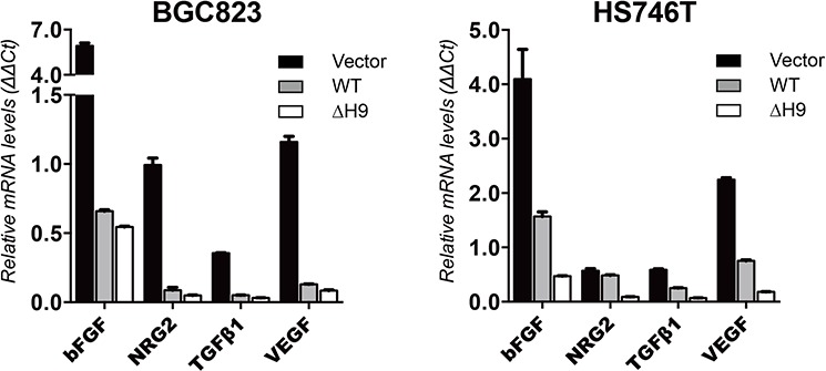
HOXB9, a transcription factor, plays an important role in development. While HOXB9 has been implicated in tumorigenesis and metastasis, its mechanisms are variable and its role in gastric carcinoma (GC) remains unclear. In the present study, we demonstrated that the expression of HOXB9 decreased in gastric carcinoma and was associated with malignancy and metastasis. Re-expression of HOXB9 in gastric cell lines resulted in the suppression of cell proliferation, migration, and invasion, which was accompanied by the induction of mesenchymal-to-epithelial transition (MET). Comparative sequence analysis and examination of a HOXB9 structural model indicated that three sites might possibly be involved in MET regulation. The in vitro study of HOXB9 mutants showed that these were unable to inhibit MET induction. However, when overexpressing a HOXB9 mutant lacking the hexapeptide motif, a more potent MET induction and tumor suppression was observed compared to that of the wild-type, indicating that the presence of the hexapeptide motif reduced HOXB9 MET induction and tumor suppression activity. Therefore, the results of the present study suggested that HOXB9 is a tumor suppressor in gastric carcinoma, and its activity was controlled by different regulatory mechanisms such as the hexapeptide motif as a "brake" in this case. The results of these regulatory effects could lead to either oncogenic or tumor suppressive roles of HOXB9, depending on the context of the particular type of cancer involved.

Keywords: HOXB9; gastric carcinoma; hexapeptide; mesenchymal-to-epithelial transition.


Conflict of interest statement

CONFLICTS OF INTEREST

The authors declare no conflict of interest.

Figures

Figure 1
Figure 1. Immunohistochemical staining of HOXB9 in gastric tissues
A. In normal tissues adjacent to a gastric adenocarcinoma, positive HOXB9 staining enriched in the epithelial cells of gastric glands. B. In intestinal-type gastric adenocarcinoma tissues, decreased expression of HOXB9 in cancer cells. C. In diffuse-type gastric adenocarcinoma tissues, no discernable staining of HOXB9. Original magnification was 10× in all photomicrographs.
Figure 2
Figure 2. HOXB9 suppressed multiple malignant phenotypes of gastric carcinoma in vitro through a mesenchymal-to-epithelial transition (MET)
BGC823 and HS746T cells were transfected with HOXB9 or a non-targeting control and checked with A. Cell Counting Kit-8 assays, B. colony formation assays, C. Transwell® migration and invasion assays, D. wound healing cell migration assays, and E. western blot to detect MET markers such as E-cadherin, N-cadherin, Snail and Vimentin. Bars indicate standard errors (n = 5, P < 0.05).
Figure 3
Figure 3. Comparative sequence analysis of HOXB9
A. The arrangement of HoxB cluster on human chromosome 17 (ch17). HOXB9 is positioned in the 5′ cluster as a conventional posterior Hox gene. B. Sequence alignment of homeodomains (green) and hexapeptide motifs (red) within the HoxB cluster and Hox9 paralog proteins. Residues Pro191, Tyr192 and Thr197 are DNA mediation residues and are highlighted in yellow. DNA recognition residues are colored ochre. The predicted secondary structure is shown on the top and colored in accordance with the aligned residues. C. The distribution of HOXB9 mutations that were identified in various tumors and are predicted to result in HOXB9 amino acid substitutions. The data was summarized according to the Catalogue of Somatic Mutations In Cancer (COSMIC) database. The mutations identified in gastric carcinomas are highlighted in red, and the truncating mutations are indicated by the gray shadow.
Figure 4
Figure 4. The structural models of HOXB9
A. A tertiary structure of HOXB9 predicted by Swiss-Model. The homeodomain is shown in green, the hexapeptide motif in red and a linker in gray. The N-terminal flexible region, which cannot build a model, is shown as a purple dashed line. B. Validation of HOXB9 model using a Ramachandran plot. Plot statistics show that 94.3% of the residues were in most favored regions (A, B, L), 5.7% were in additional allowed regions (a, b, l, p) and 0% in disallowed regions. Usually, a good quality model would have > 90% residues in the most favored regions. The results were based on the analysis of 118 structures, with a resolution of >2.0 Angstroms and R-factor no greater than 20%. C. A model mimicking HOXB9 interacting with double-strand DNA and Pbx1 based on the crystal structure of HOXA9-Pbx1-DNA complex (PDB: 1PUF). The HOXB9 model is depicted in green, the DNA in yellow and Pbx1 in light blue. The HOXB9 hexapeptide motif, which interacts with Pbx1 via the Trp179 residue, is depicted in red. The N-terminal flexible region, which interacts with Btg1 or Btg2, is invisible in the model and is depicted as a purple dashed line. In the homeodomain, arrowheads indicate the location of DNA recognition residues, and arrows indicate DNA mediation residues. D. A schematic view of wild-type (WT) HOXB9, the hexapeptide motif deletion mutant (ΔH9), the DNA mediation residues substitution mutant (Tri-mu), and the N-terminal 1–78 amino acids deletion mutant (ΔN78).
Figure 5
Figure 5. Analysis of the potential regulatory sites in HOXB9
BGC823 and HS746T cells were transfected with WT, ΔN78, Tri-mu, ΔH9 of HOXB9, or the non-targeting control. The expression of mesenchymal-to-epithelial transition (MET) markers such as E-cadherin, N-cadherin, Snail, and Vimentin was detected via western blot A. The expression of MET markers in HS746T cells after transfection was quantified with normalization to GAPDH. Bars indicate standard errors (n = 3) B. The malignant phenotypes of BGC823 and HS746T cells after transfection with WT or ΔH9 HOXB9 or the non-targeting control were analyzed with Cell Counting Kit-8 assays for cell proliferation C. and Transwell® migration and invasion assays for migration and invasion D. Bars indicate standard errors (n = 5, P < 0.05).
Figure 6
Figure 6. Immunocytochemistry analysis of cell morphogenesis during MET induction in gastric carcinoma cells
HS746T cells were transfected with WT or ΔH9 of HOXB9 or the non-targeting control and stained with the E-cadherin (green) and Vimentin (red) antibodies, and the nuclei counterstained with DAPI (blue). Three figures were merged to observe E-cadherin/Vimentin ratio changes as well as morphological changes (Magnification: 40× ; Magnification of yellow box: 100×). Bars indicate 100 μm.
Figure 7
Figure 7. The expression of HOXB9 downstream effective genes in GC cells
The BGC823 and HS746T cells were transfected with vector only, WT, and ΔH9 HOXB9. The relative mRNA level expression of bFGF, NRG2, TGF-β, and VEGF in these cells were quantified using real-time PCR and normalized against GAPDH. Bars indicate standard errors (n = 3).
Figure 8
Figure 8. Schematic model of the restricted MET induction in gastric carcinoma (GC) cells by HOXB9
HOXB9 suppressed malignancy and metastasis of GC cells by inducing MET, which was mediated by specific regulatory sites in the HOXB9 protein. However, MET induction by HOXB9 was negatively regulated by the hexapeptide motif (indicated by a solid line), which may be through the interaction with its cofactors such as Pbx1, and the GC cells may actually follow the direction of EMT. When the hexapeptide motif was impaired (indicated by a dashed line), HOXB9 induced a higher MET rate in the cells. MET: mesenchymal-to-epithelial transition, EMT: epithelial-to-mesenchymal transition.

References

    1. Gehring WJ, Affolter M, Burglin T. Homeodomain proteins. Annual review of biochemistry. 1994;63:487–526. - PubMed
    1. Catron KM, Iler N, Abate C. Nucleotides flanking a conserved TAAT core dictate the DNA binding specificity of three murine homeodomain proteins. Molecular and cellular biology. 1993;13:2354–2365. - PMC - PubMed
    1. Apiou F, Flagiello D, Cillo C, Malfoy B, Poupon MF, Dutrillaux B. Fine mapping of human HOX gene clusters. Cytogenetics and cell genetics. 1996;73:114–115. - PubMed
    1. Lewis EB. A gene complex controlling segmentation in Drosophila. Nature. 1978;276:565–570. - PubMed
    1. Garcia-Fernandez J. The genesis and evolution of homeobox gene clusters. Nature reviews Genetics. 2005;6:881–892. - PubMed

Publication types

MeSH terms