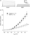
BDNF contributes to angiotensin II-mediated reductions in peak voltage-gated K+ current in cultured CATH.a cells
- PMID: 26537343
- PMCID: PMC4673628
- DOI: 10.14814/phy2.12598
BDNF contributes to angiotensin II-mediated reductions in peak voltage-gated K+ current in cultured CATH.a cells
Abstract
Increased central angiotensin II (Ang II) levels contribute to sympathoexcitation in cardiovascular disease states such as chronic heart failure and hypertension. One mechanism by which Ang II increases neuronal excitability is through a decrease in voltage-gated, rapidly inactivating K(+) current (IA); however, little is known about how Ang II signaling results in reduced IA. Brain-derived neurotrophic factor (BDNF) has also been demonstrated to decrease IA and has signaling components common to Ang II. Therefore, we hypothesized that Ang II-mediated suppression of voltage-gated K(+) currents is due, in part, to BDNF signaling. Differentiated CATH.a, catecholaminergic cell line treated with BDNF for 2 h exhibited a reduced IA in a manner similar to that of Ang II treatment as demonstrated by whole-cell patch-clamp analysis. Inhibiting BDNF signaling by pretreating neurons with an antibody against BDNF significantly attenuated the Ang II-induced reduction of IA. Inhibition of a common component of both BDNF and Ang II signaling, p38 MAPK, with SB-203580 attenuated the BDNF-mediated reductions in IA. These results implicate the involvement of BDNF signaling in Ang II-induced reductions of IA, which may cause increases in neuronal sensitivity and excitability. We therefore propose that BDNF may be a necessary component of the mechanism by which Ang II reduces IA in CATH.a cells.
Keywords: AT1R; SB‐203580; TrkB; p38 MAPK.
© 2015 The Authors. Physiological Reports published by Wiley Periodicals, Inc. on behalf of the American Physiological Society and The Physiological Society.
Figures


References
-
- Chan SHH, Wu C-WJ, Chang AYW, Hsu K-S. Chan JYH. Transcriptional upregulation of brain-derived neurotrophic factor in rostral ventrolateral medulla by angiotensin II: significance in superoxide homeostasis and neural regulation of arterial pressure. Circ. Res. 2010;107:1127–1139. - PubMed
Grants and funding
LinkOut - more resources
Full Text Sources
Other Literature Sources
Miscellaneous

