Review
. 2016 Jan;96(1):1-17.
doi: 10.1152/physrev.00013.2015.

Spermatogenesis: The Commitment to Meiosis

Affiliations
Review

Spermatogenesis: The Commitment to Meiosis

Michael D Griswold. Physiol Rev. 2016 Jan.

Abstract

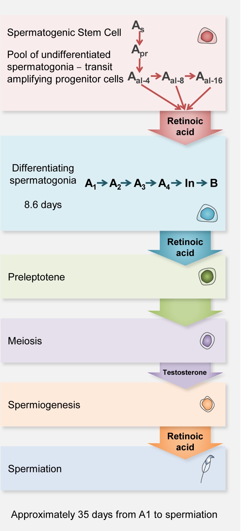
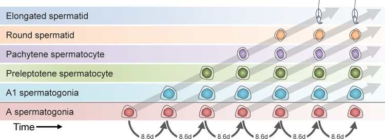
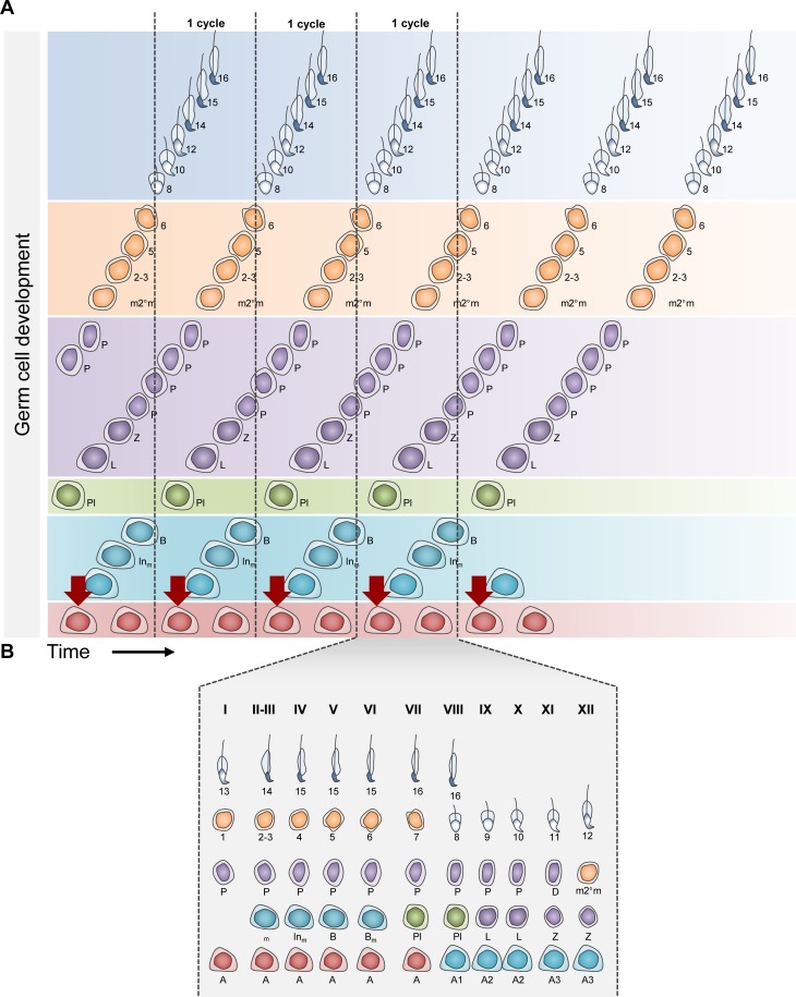
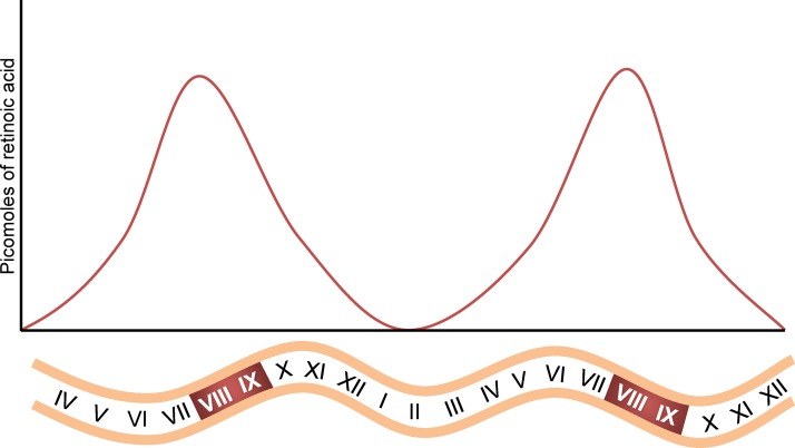
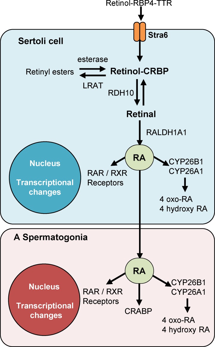
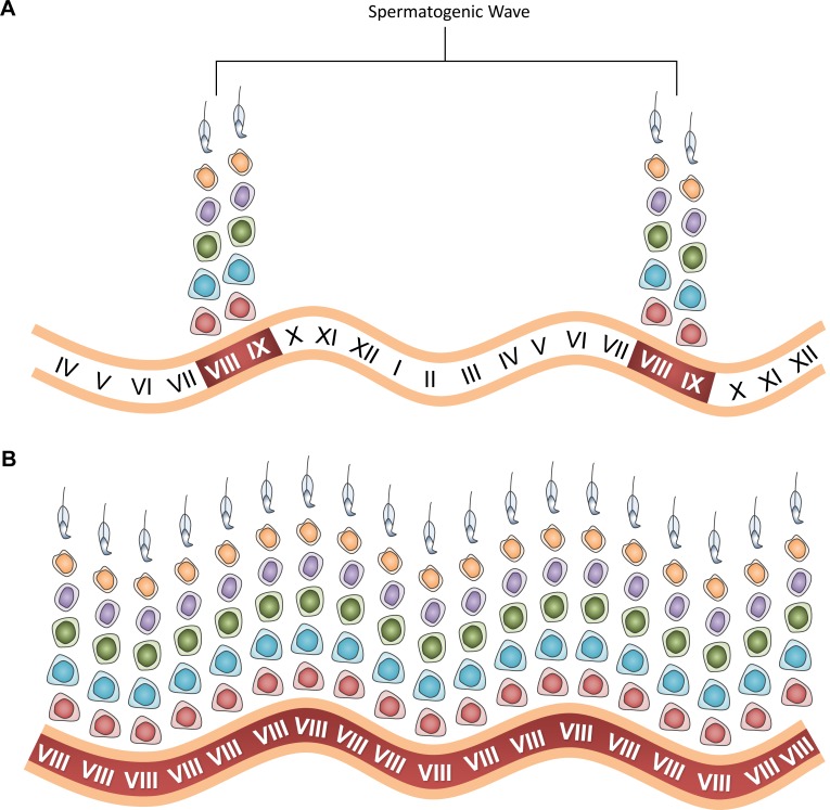
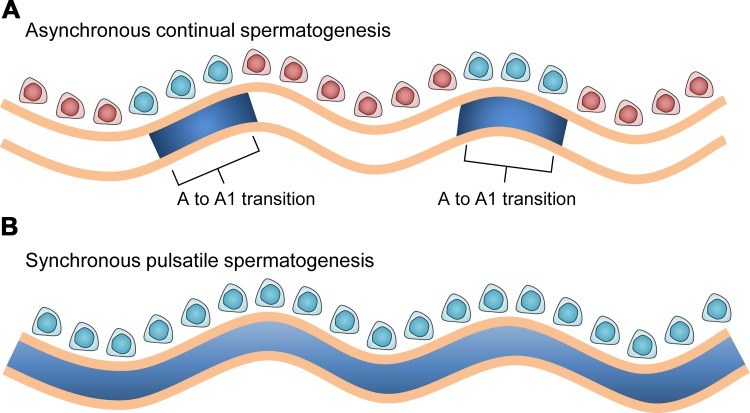
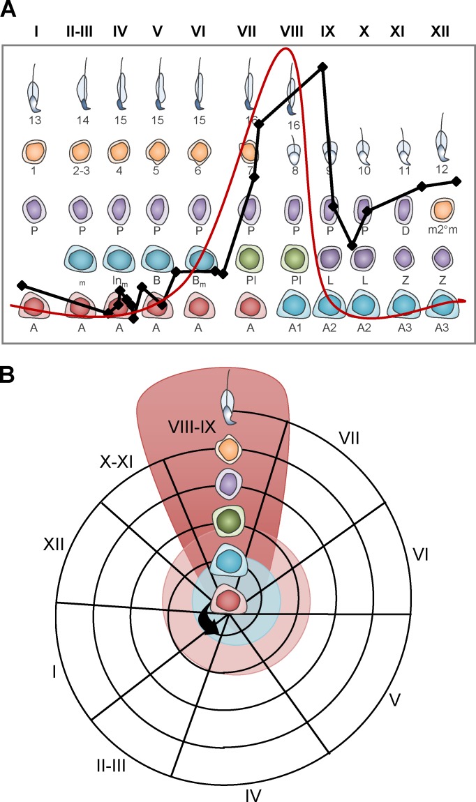
Mammalian spermatogenesis requires a stem cell pool, a period of amplification of cell numbers, the completion of reduction division to haploid cells (meiosis), and the morphological transformation of the haploid cells into spermatozoa (spermiogenesis). The net result of these processes is the production of massive numbers of spermatozoa over the reproductive lifetime of the animal. One study that utilized homogenization-resistant spermatids as the standard determined that human daily sperm production (dsp) was at 45 million per day per testis (60). For each human that means ∼1,000 sperm are produced per second. A key to this level of gamete production is the organization and architecture of the mammalian testes that results in continuous sperm production. The seemingly complex repetitious relationship of cells termed the "cycle of the seminiferous epithelium" is driven by the continuous commitment of undifferentiated spermatogonia to meiosis and the period of time required to form spermatozoa. This commitment termed the A to A1 transition requires the action of retinoic acid (RA) on the undifferentiated spermatogonia or prospermatogonia. In stages VII to IX of the cycle of the seminiferous epithelium, Sertoli cells and germ cells are influenced by pulses of RA. These pulses of RA move along the seminiferous tubules coincident with the spermatogenic wave, presumably undergoing constant synthesis and degradation. The RA pulse then serves as a trigger to commit undifferentiated progenitor cells to the rigidly timed pathway into meiosis and spermatid differentiation.


Figures

Figure 1.
Figure 1.
Overview of germ cells differentiation in mice. After the initial round where differentiating spermatogonia develop directly from prospermatogonia, the subsequent rounds of cells arise from a subset of A single spermatogonia (As) termed spermatogenic stem cells (SSC). The SSCs can divide to form A paired spermatogonia (Apr) that divide randomly through the cycle of the seminiferous epithelium stages X to VII to form A aligned cell syncytia of 4, 8, and 16 cells. This pool of ”undifferentiated“ spermatogonia (red cell) have also been termed ”transit amplifying progenitor cells," and nearly all (with exception of the SSCs) transition without cell division into A1 differentiating spermatogonia. The differentiating spermatogonia (teal cell) undergo 5 cell divisions synchronized to the cycle of the seminiferous epithelium to form B spermatogonia. Another mitosis results in the formation of preleptotene spermatocytes (green cell). The preleptotene spermatocytes proceed through the rest of meiosis (purple cell), form haploid round or elongating spermatids (orange cell) and eventually elongated spermatids (blue cell). This color key is maintained for the next few figures. Androgen receptor gene deletion studies demonstrate the requirement of testosterone for early-stage elongating spermatids, terminal differentiation, and release of spermatids (56).
Figure 2.
Figure 2.
Generating the recurring cellular associations that define the cycle of the seminiferous epithelium. The cycle is generated by the precisely timed transition of A spermatogonia (red) into A1 spermatogonia (teal). In the mouse this transition occurs every 8.6 days. In addition, it takes 8.6 days for the A1 spermatogonia to become preleptotene spermatocytes (green) and enter meiosis and an additional 8.6 days ×3 to form elongated spermatids ready for spermiation. The net result is that once fully established the same cell associations or the same group of cell types appear every 8.6 days. In the 8.6-day period between the transition of A spermatogonia to A1 spermatogonia, there is a continuum of development of each cell type. Red, undifferentiated A spermatogonia; blue, differentiating A1 spermatogonia; green, preleptotene spermatocytes; purple, pachytene spermatocytes; orange, round or elongating spermatids; blue, elongated spermatids.
Figure 3.
Figure 3.
The cycle of the seminiferous epithelium and how it is derived. This figure expands on Figure 2 and utilizes the same color code for different germ cells. In A, the developmental steps from the A spermatogonia transition to A1 spermatogonia (red arrow) is shown in more detail, and several complete cycles are shown (rectangles outlined by dotted lines). Each rectangle represents 1 complete repeat of the cellular associations. In B, one rectangle is expanded to show the cycle of the seminiferous epithelium for the mouse.
Figure 4.
Figure 4.
Proposed pulses of retinoic acid along the tubule during the spermatogenic wave. The red shading represents the proposed retinoic acid pulse that drives the retinoic acid responsive events during the cycle of the seminiferous epithelium at late stage VII as well as stages VIII and IX. The relatively high concentration of retinoic acid in these stages was proposed to drive the A spermatogonia to A1 spermatogonia transition specifically at this point in the cycle.
Figure 5.
Figure 5.
Possible pathway for the flow of retinoids during the initiation of spermatogenesis. In general, the original source of RA to drive the differentiation of spermatogonia in the initial rounds appears to be the Sertoli cells where retinol is oxidized to retinal by RDH10 and then to RA by RALDH1a1. The cellular localizations of the enzymes and binding proteins are based on immunocytochemistry and expression arrays of RiboTag mice (34). Retinol-RBP4-TTR, retinol bound to the retinol binding protein 4, transthyretin complex; STRA6, stimulated by retinoic acid gene 6 cell membrane receptor; LRAT, lecithin retinol transferase; CRBP, cellular retinol binding protein; RDH10, retinol dehydrogenase 10; RALDH1a1, retinaldehyde dehydrogenase 1a1; RA, retinoic acid; CYP26, cytochrome P-450 enzymes from the cyp26 family; RAR, retinoic acid receptor; RXR, rexinoid receptor; CRABP, cellular retinoic acid receptor; RBP4, retinol binding protein 4.
Figure 6.
Figure 6.
Illustration of asynchronous versus synchronous spermatogenesis. In normal asynchronous spermatogenesis (A), a wave is generated as the retinoic acid pulses (red patches) move along the tubules driving the A to A1 transition of spermatogonia. Only stages VIII and IX are shown. In synchronous spermatogenesis (B), no wave is generated and the entire testis moves through the cellular associations at the same time. RA concentration would peak in stages VII–IX in the entire testis, and sperm would be released from the entire testis only every 8.6 days. Red, undifferentiated A spermatogonia; teal, differentiating A1 spermatogonia; green, preleptotene spermatocytes; purple, pachytene spermatocytes; orange, round or elongating spermatids; blue, elongated spermatids.
Figure 7.
Figure 7.
Initiation of the cycle of the seminiferous epithelium and spermatogenic wave as demonstrated using the RARE-hsplac mice that express β-galactosidase when an intact RA signaling system and sufficient ligand are present (99). A and B illustrate a seminiferous tubule from the RARE-hsplac mice showing areas of active retinoic acid signaling in blue. In A, the tubule from the untreated mice shows patchy areas of blue that are the harbingers of the asynchronous spermatogenesis in the adult. In B, the tubule from the mice treated with RA does not have a patchy appearance but has undergone the A to A1 transition along the entire length of the tubule. In the adult, these mice would have synchronous spermatogenesis. Red, undifferentiated spermatogonia; teal, differentiating spermatogonia.
Figure 8.
Figure 8.
Measurement of the retinoic acid pulse. The red curve (A) illustrates a hypothetical pulse of RA based on the immunocytochemistry and testicular response to RA as described in the text. The black curve is a representation of the data obtained from actual measurement of RA levels in stage synchronized testes (51). See Hogarth et al. (51) for details of the methods and actual values for the RA levels. In B, an alternative depiction of the cycle of the seminiferous epithelium is presented. The cycle begins with the undifferentiated spermatogonia (red cell) undergoing the transition to differentiating spermatogonia (teal cell). The black arrow represents the direction for progression of differentiation. The stages are shown as part of the helical wheel, and the area of the cycle where the RA peak is shown has a dark red background. The regions of spermatogonial development corresponding to undifferentiated spermatogonia (light blue oval) and the differentiating spermatogonia (pink oval) are as shown in Figure 1.

References

    1. Amann R. The cycle of the seminiferous epithelium in humans: a need to revisit? J Androl 29: 469–487, 2008. - PubMed
    1. Anderson EL, Baltus AE, Roepers-Gajadien HL, Hassold TJ, de Rooij DG, van Pelt AM, Page DC. Stra8 and its inducer, retinoic acid, regulate meiotic initiation in both spermatogenesis and oogenesis in mice. Proc Natl Acad Sci USA 105: 14976–14980, 2008. - PMC - PubMed
    1. Arnold SL, Kent T, Hogarth CA, Schlatt S, Prasad B, Haenisch M, Walsh T, Muller CH, Griswold MD, Amory JK, Isoherranen N. Importance of ALDH1A enzymes in determining human testicular retinoic acid concentrations. J Lipid Res 56: 342–357, 2015. - PMC - PubMed
    1. Ballow D, Meistrich ML, Matzuk M, Rajkovic A. Sohlh1 is essential for spermatogonial differentiation. Dev Biol 294: 161–167, 2006. - PubMed
    1. Baltus AE, Menke DB, Hu YC, Goodheart ML, Carpenter AE, de Rooij DG, Page DC. In germ cells of mouse embryonic ovaries, the decision to enter meiosis precedes premeiotic DNA replication. Nat Genet 38: 1430–1434, 2006. - PubMed

Publication types