Review
. 2015 Sep:45:48-56.
doi: 10.1016/j.semcdb.2015.10.026. Epub 2015 Oct 30.

Membrane fusion in muscle development and repair

Affiliations
Review

Membrane fusion in muscle development and repair

Alexis R Demonbreun et al. Semin Cell Dev Biol. 2015 Sep.

Abstract

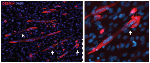
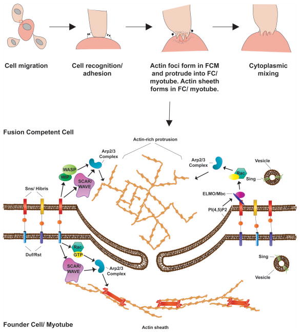
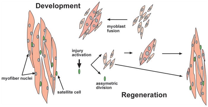
Mature skeletal muscle forms from the fusion of skeletal muscle precursor cells, myoblasts. Myoblasts fuse to other myoblasts to generate multinucleate myotubes during myogenesis, and myoblasts also fuse to other myotubes during muscle growth and repair. Proteins within myoblasts and myotubes regulate complex processes such as elongation, migration, cell adherence, cytoskeletal reorganization, membrane coalescence, and ultimately fusion. Recent studies have identified cell surface proteins, intracellular proteins, and extracellular signaling molecules required for the proper fusion of muscle. Many proteins that actively participate in myoblast fusion also coordinate membrane repair. Here we will review mammalian membrane fusion with specific attention to proteins that mediate myoblast fusion and muscle repair.

Keywords: Development; Fusion; Membrane; Muscle; Myoblast; Repair.


Conflict of interest statement

Conflict of Interest

There are no conflicts of interest.

Figures

Fig. 1
Fig. 1
Membranes in muscle. Mature muscle is composed of individual multinucleate myofibers bound by plasma membrane, or sarcolemma. The sarcoplasmic reticulum (SR) is specialized endoplasmic reticulum, and the transverse (T)-tubules are sarcolemmal invaginations evolved for Ca2+ handling and contraction. The SR forms a complex network around the myofiber adjacent to the T-tubules. Muscle striations are generated from the thin-filament (actin) containing I-band and the thick-filament (myosin) containing A-band. The Z-disk defines the edges of the sarcomere. Muscle contraction occurs as myosin slides across the filaments shortening the sarcomere.
Fig. 2
Fig. 2
Myoblast fusion in mammals. During muscle growth satellite cells (green) give rise to mono-nucleated muscle precursor cells, myoblasts (light pink). Myoblasts fuse with one-another to generate nascent myotubes. During later steps of development, myoblasts fuse with existing myotubes promoting muscle growth. Upon injury, satellite cells are activated and asymmetrically divide generating a new pool of myoblasts. These myoblasts once again fuse with each other and to injured myotubes to promote muscle regeneration.
Fig. 3
Fig. 3
Myoblast fusion in Drosophila. Two populations of myoblasts exist during Drosophila myoblast fusion: the founder cells (FCs) and the fusion competent myoblasts (FCMs). The FCs are present at sites where mature muscle will eventually form. FCMs migrate to the FCs, adapting a characteristic teardrop form. Adhesion molecules on the cell surface of both FCs and FCMs mediate the initial recognition/adhesion step. Next, the FCM form actin rich podosome-like strictures, which extend into the FCs, while the FCs form a thin actin sheath at the membrane. Tiny pores form and expand allowing cytoplasmic mixing, and ultimately the fusion of myoblasts, resulting in a multinucleated muscle fiber. Importantly, FCMs continue to fuse to this multinucleated fiber, resulting in mature muscle.
Fig. 4
Fig. 4
Cell culture model of myoblast fusion. Primary myoblasts will differentiate and fuse with serum withdrawal. Desmin (red) is used to mark committed muscle cells. Mono-nucleated myoblasts (arrow) fuse with existing myotubes (arrowhead). The right panel shows a higher magnification view of the dotted box showing the fusion of a myoblast to an existing multinucleate myotube. DAPI (blue) stains nuclei.

References

    1. Franzini-Armstrong C, Porter KR. Sarcolemmal Invaginations Constituting the T System in Fish Muscle Fibers. The Journal of cell biology. 1964;22:675–96. - PMC - PubMed
    1. Campbell KP, Stull JT. Skeletal muscle basement membrane-sarcolemma-cytoskeleton interaction minireview series. The Journal of biological chemistry. 2003;278:12599–600. - PubMed
    1. Yin H, Price F, Rudnicki MA. Satellite cells and the muscle stem cell niche. Physiological reviews. 2013;93:23–67. - PMC - PubMed
    1. Cooper ST, Head SI. Membrane Injury and Repair in the Muscular Dystrophies. Neuroscientist. 2014 - PubMed
    1. Abmayr SM, Pavlath GK. Myoblast fusion: lessons from flies and mice. Development. 2012;139:641–56. - PMC - PubMed

Publication types

Substances