

Structural Studies of Chikungunya Virus-Like Particles Complexed with Human Antibodies: Neutralization and Cell-to-Cell Transmission
- PMID: 26537684
- PMCID: PMC4719628
- DOI: 10.1128/JVI.02364-15
Structural Studies of Chikungunya Virus-Like Particles Complexed with Human Antibodies: Neutralization and Cell-to-Cell Transmission
Abstract
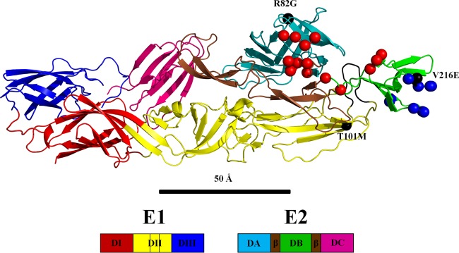
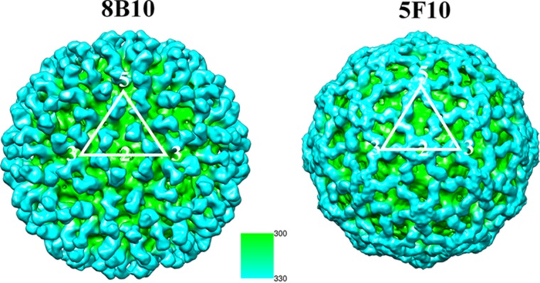
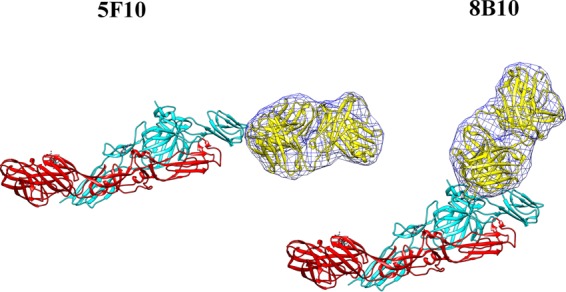
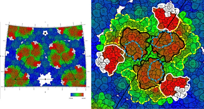
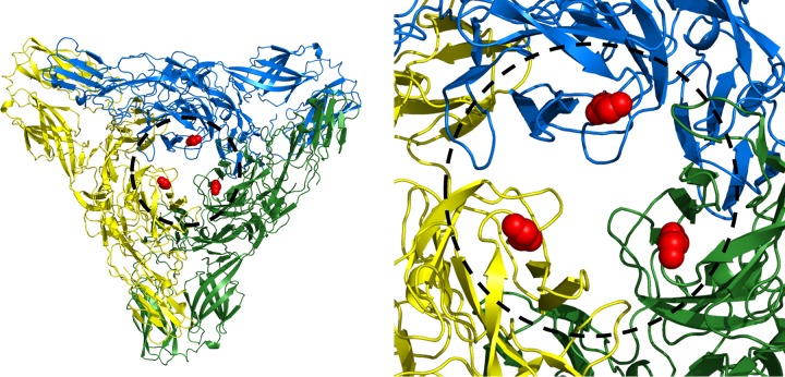
Chikungunya virus is a positive-stranded RNA alphavirus. Structures of chikungunya virus-like particles in complex with strongly neutralizing antibody Fab fragments (8B10 and 5F10) were determined using cryo-electron microscopy and X-ray crystallography. By fitting the crystallographically determined structures of these Fab fragments into the cryo-electron density maps, we show that Fab fragments of antibody 8B10 extend radially from the viral surface and block receptor binding on the E2 glycoprotein. In contrast, Fab fragments of antibody 5F10 bind the tip of the E2 B domain and lie tangentially on the viral surface. Fab 5F10 fixes the B domain rigidly to the surface of the virus, blocking exposure of the fusion loop on glycoprotein E1 and therefore preventing the virus from becoming fusogenic. Although Fab 5F10 can neutralize the wild-type virus, it can also bind to a mutant virus without inhibiting fusion or attachment. Although the mutant virus is no longer able to propagate by extracellular budding, it can, however, enter the next cell by traveling through junctional complexes without being intercepted by a neutralizing antibody to the wild-type virus, thus clarifying how cell-to-cell transmission can occur.
Importance: Alphaviral infections are transmitted mainly by mosquitoes. Chikungunya virus (CHIKV), which belongs to the Alphavirus genus, has a wide distribution in the Old World that has expanded in recent years into the Americas. There are currently no vaccines or drugs against alphaviral infections. Therefore, a better understanding of CHIKV and its associated neutralizing antibodies will aid in the development of effective treatments.
Copyright © 2016, American Society for Microbiology. All Rights Reserved.
Figures
References
-
- Paquet C, Quatresous I, Solet JL, Sissoko D, Renault P, Pierre V, Cordel H, Lassalle C, Thiria J, Zeller H, Schuffnecker I. 2006. Chikungunya outbreak in Reunion: epidemiology and surveillance, 2005 to early January 2006. Euro Surveill 11:2891 http://www.eurosurveillance.org/ViewArticle.aspx?ArticleId=2891. - PubMed
-
- Kostyuchenko VA, Jakana J, Liu X, Haddow AD, Aung M, Weaver SC, Chiu W, Lok SM. 2011. The structure of Barmah Forest virus as revealed by cryo-electron microscopy at a 6-angstrom resolution has detailed transmembrane protein architecture and interactions. J Virol 85:9327–9333. doi:10.1128/JVI.05015-11. - DOI - PMC - PubMed
Publication types
MeSH terms
Substances
Associated data
- Actions
- Actions
- Actions
Grants and funding
LinkOut - more resources
Full Text Sources
Other Literature Sources
Medical
