. 2016 Feb;98(2):172-85.
doi: 10.1007/s00223-015-0076-4. Epub 2015 Nov 5.

Sex-Linked Skeletal Phenotype of Lysyl Oxidase Like-1 Mutant Mice

Affiliations

Sex-Linked Skeletal Phenotype of Lysyl Oxidase Like-1 Mutant Mice

Loai Alsofi et al. Calcif Tissue Int. 2016 Feb.

Abstract

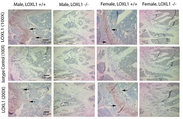
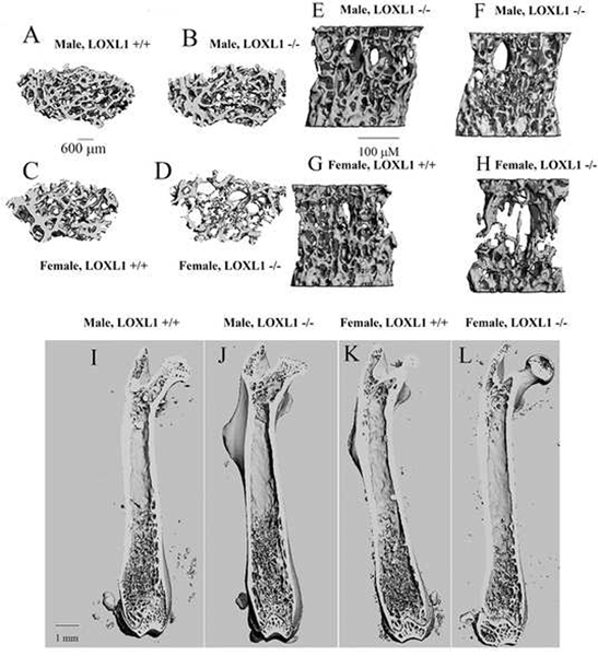
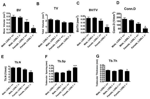
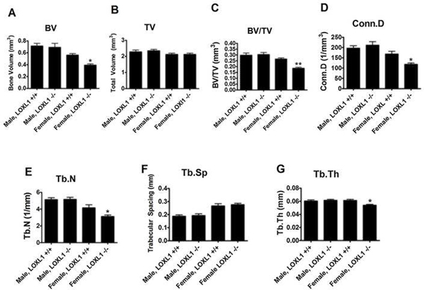
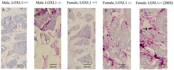
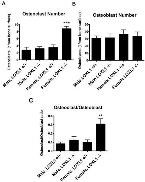
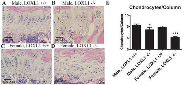
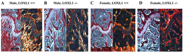
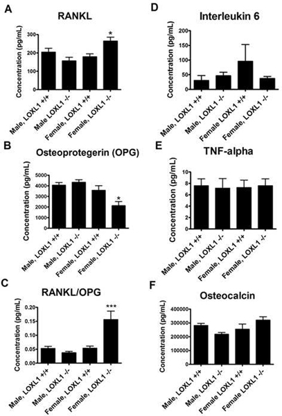
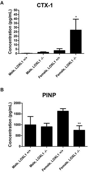
Lysyl oxidases are required for collagen and elastin cross-linking and extracellular matrix maturation including in bone. The lysyl oxidase family consists of lysyl oxidase (LOX) and 4 isoforms (LOXL1-4). Here we investigate whether deletion of LOXL1, which has been linked primarily to elastin maturation, leads to skeletal abnormalities. Left femurs (n = 8), L5 vertebrae (n = 8), and tibiae (n = 8) were analyzed by micro-computed tomography in 13-week-old wild-type (WT) and LOXL1-/- male and female mice. Right femurs (n = 8) were subjected to immunohistochemistry for LOXL1, and histochemical/histology analyses of osteoclasts and growth plates. Sera from all mice were analyzed for bone turnover markers. Results indicate strong expression of LOXL1 in wild-type growth plates in femurs. Significant deterioration of trabecular bone structure in long bones and vertebrae from female was observed but not from male, mutant mice compared with WT. Decreases in BV/TV, Conn.D, trabecular thickness, and number in the femoral distal metaphysis were observed in female, but not in male, mutant mice. Trabecular spacing was increased significantly in femurs of female mutant mice. Findings were similar in trabeculae of L5 vertebrae from female mutant mice. The number of TRAP positive osteoclasts at the trabecular bone surface was increased in female mutant mice compared with WT females, consistent with increased serum RANKL and decreased OPG levels. Analysis of bone turnover markers confirmed increased bone resorption as indicated by significantly elevated CTX-1 in the serum of female LOXL1-/- mice compared to their wild-type counterparts, as well as decreased bone formation as measured by decreased serum levels of PINP. Picrosirius red staining revealed a loss of heterogeneity in collagen organization in female LOXL1-/- mice only, with little to no yellow and orange birefringence. Organization was also impaired in chondrocyte columns in both female and male LOXL1-/- mice, but to a greater extent in females. Data indicate that LOXL1-/- mutant mice develop appendicular and axial skeletal phenotypes characterized by decreased bone volume fraction and compromised trabecular microstructure, predominantly in females.

Keywords: Bone histomorphometry; Collagen; Genetic animal model; Lysyl oxidases; Micro-comuted tomography.


Conflict of interest statement

Conflicts of Interest

All authors declare no conflicts of interest.

Figures

Figure 1.
Figure 1.. Absence of LOXL1 in femur tissue from Loxl1 −/− mice.
Genotyping of mouse tail tip DNA shows the presence of a single 175 bp band corresponding to the mutant gene and the absence of the larger band corresponding to the wild type LOXL1 gene. Genotype designations are under each lane. The marker is a 1 Kbp ladder.
Figure 2.
Figure 2.. Absence of LOXL1 in LOXL1−/− bones.
LOXL1 antibody (Abbiotec #252215) or IgG isotype control were employed to stain femurs from LOXL1−/− mice and wild type littermates. Arrows mark the growth plates which contain high in LOXL1 staining in wild type mice. LOXL1−/− femurs contain no LOXL1 staining. Scale bars, 100 μm for 100X magnification and 50 μm for 200X magnification.
Figure 3.
Figure 3.. Micro-computed tomography of Loxl1−/− and wild type mouse bones.
Three-dimensional images (transverse view) for the femoral distal metaphysis (A-D) representing wild type and LOXL1 −/− male and female mice. Images are representative of eight animals analyzed. (E-H) Images for L5 vertebrae (sagittal view) representing wild-type and LOXL1 −/− male and female mice. Images are representative of eight animals analyzed per group. (I-L) Representative full-length, cross-sectional views of the femur from wild type and LOXL1 −/− male and female mice showing the same overall length of femurs, but trabecular deficiencies in female LOXL1−/− femurs.
Figure 4.
Figure 4.. Trabecular bone parameters from femur distal metaphysis from wild type and LOXL1−/− male and female mice.
Data are (mean values +/− SD). One asterisk (*) indicates significant difference from wild-type mice of the same sex with p<.01, two asterisks (**) indicate p<.001, and three asterisks indicate p<.0001 by one-way ANOVA and Tukey’s multiple comparison test; n=8
Figure 5.
Figure 5.. Trabecular bone parameters from L5 vertebrae representing wild type and LOXL1−/− male and female mice.
Data are (mean values +/− SD). One asterisk (*) indicates significant difference from wild-type mice of the same sex with p<.01, two asterisks (**) indicate p<.001, and three asterisks indicate p<.0001 by one-way ANOVA and Tukey’s multiple comparison test; n=8
Figure 6.
Figure 6.. TRAP staining of representative sagittal sections of femurs.
(A-D) Images are from the area directly proximal to the distal growth plates representing wild type and LOXL1−/− male and female mice; scale bar, 200 uM. (E) Panel D at higher magnification (200X). Arrows indicate TRAP positive osteoclasts. Scale bar, 100 μM.
Figure 7.
Figure 7.. (A) Histomorphometric analyses of histological sections of femur sections analyzed for number of TRAP positive osteoclasts in wild type and LOXL1−/− male and female mice.
Data are (mean values +/− SD). One asterisk (*) indicates significant difference from wild-type mice of the same sex with p<.01, two asterisks (**) indicate p<.001, and three asterisks indicate p<.0001 by one-way ANOVA and Tukey’s multiple comparison test; n=8. (B) Histomorphometric analyses of histological sections of femur sections analyzed for number of osteoblasts. Asterisk notation is the same, n=8 (C) Calculated ratio of osteoclasts to osteoblasts. Asterisk notation is the same, n=8.
Figure 8.
Figure 8.. Hematoxylin and eosin staining of representative sagittal sections of femur distal growth plates.
(A) Images are from wild type and LOXL1−/− male and female mice; scale bar, 100 μM. (B) Histomorphometric analyses of histological sections of femur sections for chondrocytes per column of wild type and mutant male and female mice. Asterisk notation is the same as in Figure 6, n=8.
Figure 9.
Figure 9.. Picrosirius red staining of representative sagital sections of femurs.
Images are from the area starting at the center of the growth plate extending ~2mm to the right from wild type and LOXL1−/− male and female mice. Scale bar, 500 μM.
Figure 10.
Figure 10.. Concentration (pg/mL) of OPG, RANKL, Interleukin 6, TNF-α, and osteocalcin in the serum of wild type and LOXL1−/− male and female mice.
Data are mean values in pg/ml +/− SD. One asterisk (*) indicates significant difference from wild-type mice of the same sex with p<.01, two asterisks (**) indicate p<.001, and three asterisks indicate p<.0001 by one-way ANOVA and Tukey’s multiple comparison test; n=8.
Figure 11.
Figure 11.. Concentration (pg/mL) of PINP and CTX-1 in the serum of wild type and LOXL1−/− male and female mice.
Data are mean values in pg/ml +/− SD. One asterisk (*) indicates significant difference from wild-type mice of the same sex with p<.05 and two asterisks (**) indicate p<.01 by one-way ANOVA and Tukey’s multiple comparison test; n=6.

References

    1. Csiszar K (2001) Lysyl oxidases: a novel multifunctional amine oxidase family. Progress in nucleic acid research and molecular biology 70:1–32 - PubMed
    1. Khosravi R, Sodek KL, Xu WP, Bais MV, Saxena D, Faibish M, Trackman PC (2014) A novel function for lysyl oxidase in pluripotent mesenchymal cell proliferation and relevance to inflammation-associated osteopenia. PloS one 9:e100669. - PMC - PubMed
    1. Perryman L, Erler JT (2014) Lysyl oxidase in cancer research. Future Oncol 10:1709–1717 - PubMed
    1. Kenyon K, Contente S, Trackman PC, Tang J, Kagan HM, Friedman RM (1991) Lysyl oxidase and rrg messenger RNA. Science 253:802. - PubMed
    1. Liu G, Daneshgari F, Li M, Lin D, Lee U, Li T, Damaser MS (2007) Bladder and urethral function in pelvic organ prolapsed lysyl oxidase like-1 knockout mice. British Journal of Urology International 100:414–418 - PubMed

Publication types

Substances

LinkOut - more resources