. 2016 Feb 1;310(3):C193-204.
doi: 10.1152/ajpcell.00248.2015. Epub 2015 Nov 4.

The vascular Ca2+-sensing receptor regulates blood vessel tone and blood pressure

Affiliations

The vascular Ca2+-sensing receptor regulates blood vessel tone and blood pressure

M Schepelmann et al. Am J Physiol Cell Physiol. .

Abstract

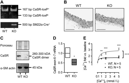
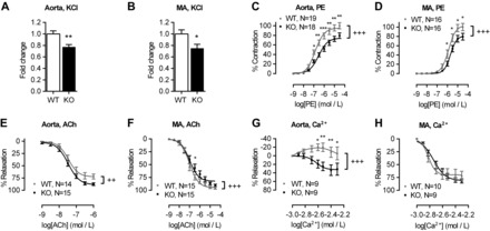
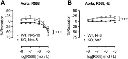
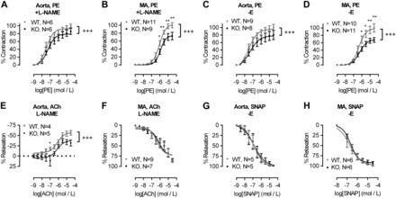
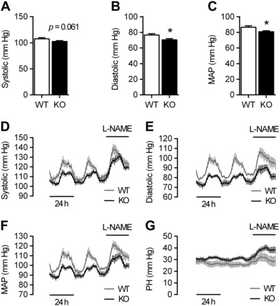
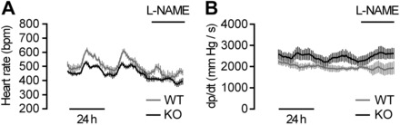
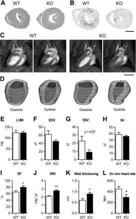
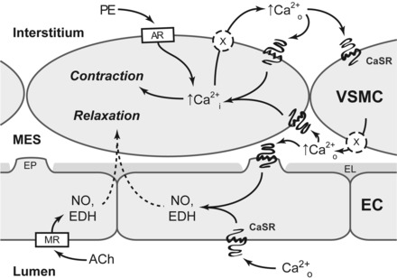
The extracellular calcium-sensing receptor CaSR is expressed in blood vessels where its role is not completely understood. In this study, we tested the hypothesis that the CaSR expressed in vascular smooth muscle cells (VSMC) is directly involved in regulation of blood pressure and blood vessel tone. Mice with targeted CaSR gene ablation from vascular smooth muscle cells (VSMC) were generated by breeding exon 7 LoxP-CaSR mice with animals in which Cre recombinase is driven by a SM22α promoter (SM22α-Cre). Wire myography performed on Cre-negative [wild-type (WT)] and Cre-positive (SM22α)CaSR(Δflox/Δflox) [knockout (KO)] mice showed an endothelium-independent reduction in aorta and mesenteric artery contractility of KO compared with WT mice in response to KCl and to phenylephrine. Increasing extracellular calcium ion (Ca(2+)) concentrations (1-5 mM) evoked contraction in WT but only relaxation in KO aortas. Accordingly, diastolic and mean arterial blood pressures of KO animals were significantly reduced compared with WT, as measured by both tail cuff and radiotelemetry. This hypotension was mostly pronounced during the animals' active phase and was not rescued by either nitric oxide-synthase inhibition with nitro-l-arginine methyl ester or by a high-salt-supplemented diet. KO animals also exhibited cardiac remodeling, bradycardia, and reduced spontaneous activity in isolated hearts and cardiomyocyte-like cells. Our findings demonstrate a role for CaSR in the cardiovascular system and suggest that physiologically relevant changes in extracellular Ca(2+) concentrations could contribute to setting blood vessel tone levels and heart rate by directly acting on the cardiovascular CaSR.

Keywords: CaSR; G protein-coupled receptor; blood pressure regulation; blood vessel tone regulation; calcium-sensing receptor; vascular smooth muscle cells.


Figures

Fig. 1.
Fig. 1.
Characterization of the SM22αCaSRΔflox/Δflox mouse. A: typical genotyping of wild-type (WT) and knockout (KO) mice. B: representative histological sections of aortas from WT and KO animals (n = 4). Scale bars = 100 μm. C: Western blot analysis of calcium-sensing receptor (CaSR) expression with Ponceau staining as loading control (top and middle) and α-smooth muscle actin (α-SM actin; bottom) expression in endothelium-denuded and adventitia-removed, pooled aortas; n = 18 (WT) and 17 (KO) mice. D: relative mRNA expression of the full-length CaSR (exon 6–7 vs. WT) in endothelium-denuded, adventitia-removed aortas of WT and KO animals; mean (line) ± 1SD (box); n = 4. **P < 0.01, Student's t-test (performed on ΔΔCt values vs. WT). E: intracellular Ca2+ concentration ([Ca2+]i) in freshly isolated WT and KO VSMC exposed to increasing extracellular Ca2+ concentration ([Ca2+]o; 0.1–5 mmol/l), reported as fold changes from baseline values. Curves were fitted as hyperbolic. Data are means ± SE; n = 3 (WT) and 4–5 (KO). **P < 0.01. ***P < 0.001, two-way ANOVA with Holm-Sidak posttest. +++P < 0.001, extra sum-of-squares F-test for curve comparison.
Fig. 2.
Fig. 2.
Blood vessel contractility of SM22αCaSRΔflox/Δflox mice is impaired. Aorta (WT, n = 33; KO, n = 30) and mesenteric artery (MA) responses to high K+ (KCl; 40 mmol/l; WT, n = 33; KO, n = 34; A and B), phenylephrine (PE; 1 nmol/l-30 μmol/l; C and D), acetylcholine (ACh, 1 nmol/l to 30 μmol/l, PE-preconstricted to ∼60% max; E and F), and Ca2+o (1–5 mmol/l, PE-preconstricted to ∼60% max; G and H). Results were normalized to the averaged maximum control response in WT. Curves were fitted as sigmoidal concentration-response (for PE and ACh) or 3rd-order polynomial (for Ca2+). Data are means ± SE. *P < 0.05, **P < 0.01, ***P < 0.001, Student's t-test, or two-way ANOVA with Holm-Sidak posttest. ++P < 0.01, +++P < 0.001, extra sum-of-squares F-test for curve comparison.
Fig. 3.
Fig. 3.
Pharmacological activation of the CaSR in WT and KO blood vessels. Responses to NPS R-568 (10 nmol/l to 10 μmol/l) in intact (A) or endothelium-denuded (B) aortas. Curves were fitted as sigmoidal concentration response. Data are means ± SE. +++P < 0.001, two-way ANOVA.
Fig. 4.
Fig. 4.
Role of nitric oxide (NO) in endothelium-dependent relaxation in WT and KO blood vessels. Aorta and MA responses to PE (1 nmol/l to 30 μmol/l) in the presence of the NO synthase inhibitor nitro-l-arginine methyl ester (l-NAME; 100 μmol/l; A and B) or endothelium denuded (-E; C and D). E and F: aorta and MA responses to ACh (1 nmol/l to 30 μmol/l) in the presence of l-NAME (100 μmol/l). G and H: relaxation of endothelium-denuded (-E) aortas and MA from WT and KO mice to increasing concentrations of the NO donor S-ntroso-N-acetylpenicillamine (SNAP). Curves were fitted as sigmoidal concentration-response. Data are means ± SE. *P < 0.05, **P < 0.01, two-way ANOVA with Holm-Sidak posttest. +++P < 0.001, extra sum-of-squares F-test for curve comparison.
Fig. 5.
Fig. 5.
Tail-cuff and radiotelemetry blood pressure measurements of WT and KO animals. AC: tail-cuff measurements of systolic (A), diastolic (B), and mean arterial pressures (MAP; C) of WT and KO mice. Data are means ± SE; n = 20 (WT) and 35 (KO). *P < 0.05, Student's t-test. D-G: longitudinal radiotelemetry measurements of systolic (D) and diastolic blood pressure (E), MAP (F), and pulse height (PH; G) of WT and KO mice in the presence or absence of l-NAME treatment. Data are means ± SE; n = 5 (WT and KO).
Fig. 6.
Fig. 6.
Longitudinal radiotelemetry measurements of heart rate (A) and the positive first derivative of the blood pressure curve (dp/dt; B) of WT and KO mice in the presence or absence of l-NAME treatment. Data are means ± SE; n = 5 (WT and KO).
Fig. 7.
Fig. 7.
Cardiac phenotype of WT and KO animals. Hematoxylin and eosin staining (A) and picrosirius red staining (B) of heart sections showing reduced occurrence of fibrosis in KO hearts compared with WT. Representative long-axis MRI scans (C) and 3D reconstructions (D) of left ventricles of WT and remodeled KO mice (5 out of 11 investigated) in end diastole and end systole. Left ventricular mass (LVM; E), left ventricular end diastolic (EDV; F), end systolic (ESV; G), and stroke volume (SV; H), ejection fraction (EF; I), diastolic remodeling index (DRI; J), and wall thickening (end-systolic minus end-diastolic wall thickness; K) of remodeled hearts. Data are means ± SE; n = 5. **P < 0.01, *P < 0.05, Student's t-test. L: base intrinsic heart rate [beats per min (bpm)] of ex vivo retrograde perfused hearts. Data are means ± SE; n = 9 (WT) and 10 (KO). *P < 0.05, Student's t-test.
Fig. 8.
Fig. 8.
Hypothetical mechanism for the CaSR mediated auto-/paracrine amplification of contraction in vascular smooth muscle cells (VSMC). In VSMC, adrenoceptor (AR) agonists (e.g., PE) increase [Ca2+i], causing blood vessel constriction. Ca2+i is then extruded from the cell into the interstitium. Locally accumulating Ca2+o activates the CaSR on the same and neighboring VSMC, thus amplifying and synchronizing VSMC contractility. Ca2+i is also extruded into the myo-endothelial space (MES) where it can activate the CaSR on endothelial cell (EC) projections (EP), which penetrate the internal elastic lamina (EL). Activation of the endothelial CaSR leads to VSMC relaxation via a mechanism likely involving NO synthesis and endothelium-derived hyperpolarizations (EDH), in a fashion similar to dilating agents like acetylcholine (ACh) acting on muscarinic receptors (MR).

References

    1. Alam MU, Kirton JP, Wilkinson FL, Towers E, Sinha S, Rouhi M, Vizard TN, Sage AP, Martin D, Ward DT, Alexander MY, Riccardi D, Canfield AE. Calcification is associated with loss of functional calcium-sensing receptor in vascular smooth muscle cells. Cardiovasc Res 81: 260–268, 2009. - PubMed
    1. Biernacka A, Frangogiannis NG. Aging and cardiac fibrosis. Aging Dis 2: 158–173, 2011. - PMC - PubMed
    1. Block GA, Martin KJ, de Francisco AL, Turner SA, Avram MM, Suranyi MG, Hercz G, Cunningham J, Abu-Alfa AK, Messa P, Coyne DW, Locatelli F, Cohen RM, Evenepoel P, Moe SM, Fournier A, Braun J, McCary LC, Zani VJ, Olson KA, Drueke TB, Goodman WG. Cinacalcet for secondary hyperparathyroidism in patients receiving hemodialysis. N Engl J Med 350: 1516–1525, 2004. - PubMed
    1. Boedtkjer E, Praetorius J, Matchkov VV, Stankevicius E, Mogensen S, Fuchtbauer AC, Simonsen U, Fuchtbauer EM, Aalkjaer C. Disruption of Na+,HCO3− cotransporter NBCn1 (slc4a7) inhibits NO-mediated vasorelaxation, smooth muscle Ca2+ sensitivity, and hypertension development in mice. Circulation 124: 1819–1829, 2011. - PubMed
    1. Broegger T, Jacobsen JC, Secher Dam V, Boedtkjer DM, Kold-Petersen H, Pedersen FS, Aalkjaer C, Matchkov VV. Bestrophin is important for the rhythmic but not the tonic contraction in rat mesenteric small arteries. Cardiovasc Res 91: 685–693, 2011. - PubMed

Publication types

MeSH terms