. 2016 May;14(5):1302-15.
doi: 10.1111/pbi.12497. Epub 2015 Nov 5.

Introducing an algal carbon-concentrating mechanism into higher plants: location and incorporation of key components

Affiliations

Introducing an algal carbon-concentrating mechanism into higher plants: location and incorporation of key components

Nicky Atkinson et al. Plant Biotechnol J. 2016 May.

Abstract

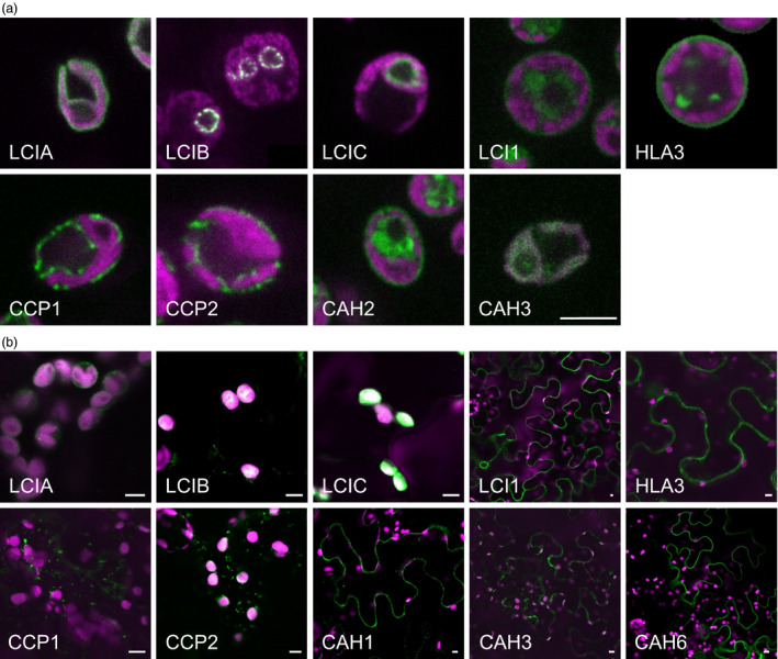
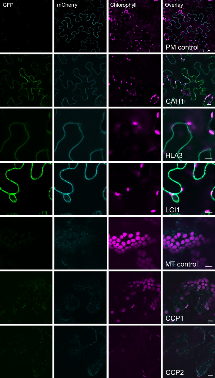
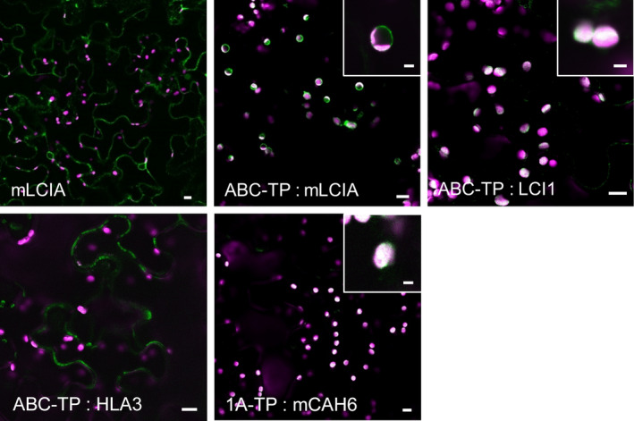
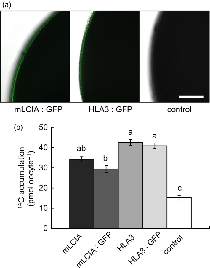
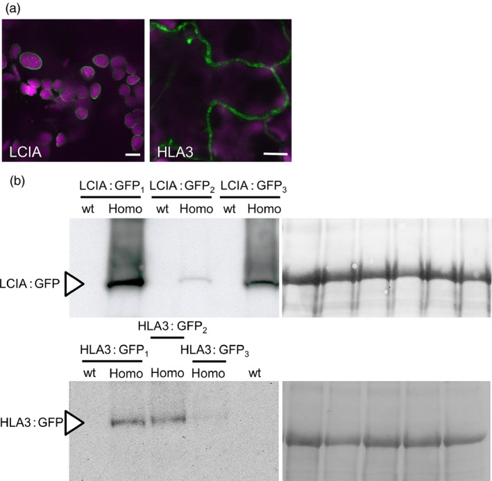
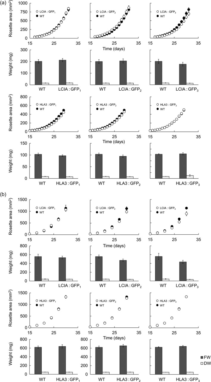
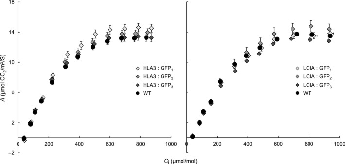
Many eukaryotic green algae possess biophysical carbon-concentrating mechanisms (CCMs) that enhance photosynthetic efficiency and thus permit high growth rates at low CO2 concentrations. They are thus an attractive option for improving productivity in higher plants. In this study, the intracellular locations of ten CCM components in the unicellular green alga Chlamydomonas reinhardtii were confirmed. When expressed in tobacco, all of these components except chloroplastic carbonic anhydrases CAH3 and CAH6 had the same intracellular locations as in Chlamydomonas. CAH6 could be directed to the chloroplast by fusion to an Arabidopsis chloroplast transit peptide. Similarly, the putative inorganic carbon (Ci) transporter LCI1 was directed to the chloroplast from its native location on the plasma membrane. CCP1 and CCP2 proteins, putative Ci transporters previously reported to be in the chloroplast envelope, localized to mitochondria in both Chlamydomonas and tobacco, suggesting that the algal CCM model requires expansion to include a role for mitochondria. For the Ci transporters LCIA and HLA3, membrane location and Ci transport capacity were confirmed by heterologous expression and H(14) CO3 (-) uptake assays in Xenopus oocytes. Both were expressed in Arabidopsis resulting in growth comparable with that of wild-type plants. We conclude that CCM components from Chlamydomonas can be expressed both transiently (in tobacco) and stably (in Arabidopsis) and retargeted to appropriate locations in higher plant cells. As expression of individual Ci transporters did not enhance Arabidopsis growth, stacking of further CCM components will probably be required to achieve a significant increase in photosynthetic efficiency in this species.

Keywords: Arabidopsis thaliana; Chlamydomonas reinhardtii; bicarbonate transporter; carbon-concentrating mechanism; photosynthesis improvement; tobacco.


Figures

Figure 1
Figure 1
Expression of fluorescent‐tagged CCM components in Chlamydomonas and tobacco. Expression of Venus‐fused CCM components in Chlamydomonas reinhardtii (a). Expression in tobacco of GFP‐fused CCM components from Chlamydomonas (b). Green and purple signals are Venus or GFP fluorescence and chlorophyll autofluorescence, respectively. Overlaid images of these signals are shown: overlaps are white. Scale bar = 5 μm (all 5 μm for Chlamydomonas images). For images of separate signals see Figure S1.
Figure 2
Figure 2
Co‐expression of GFP‐fused CCM components with a mCherry‐fused plasma membrane transporter NPSN12 or a known mitochondrial marker (the targeting sequence of yeast cytochrome oxidase IV [COX4] fused to mCherry) in tobacco. Purple, green and cyan signals are chlorophyll autofluorescence, GFP and mCherry fluorescence, respectively. Overlaid images of these signals are shown: overlaps of GFP and mCherry are pale green. PM, plasma membrane; MT, mitochondria. Scale bar = 10 μm.
Figure 3
Figure 3
Expression of GFP‐fused CCM components carrying native Arabidopsis chloroplast transit peptides in tobacco. Green and purple signals are GFP fluorescence and chlorophyll autofluorescence, respectively. Overlaid images of these signals are shown: overlaps are white. 1A‐TP, RuBisCO small subunit RBCS1A (AT1G67090) transit peptide; ABCTP, ABC transporter ABCI13 (AT1G65410) transit peptide; mCAH6, mature CAH6; mLCIA, mature LCIA. Main image scale bar = 10 μm, inset image scale bar = 3 μm. For images of separate signals see Figure S3.
Figure 4
Figure 4
Chlamydomonas CCM components LCIA and HLA3 facilitate increased accumulation of inorganic carbon in Xenopus oocytes. Confocal images of oocytes expressing GFP fused to mature LCIA (LCIA lacking a chloroplast transit peptide, mLCIA) or HLA3 3 d after injection (a). 14C accumulation in oocytes expressing mLCIA or HLA3 either untagged or fused to GFP following 10‐min incubation in MBS containing 0.12 mM NaH14 CO 3 (b). Values are means of measurements on 20 oocytes; bars are means ± standard error (SE). Letters above the bars indicate a difference or between values; where a, b and c indicate significant difference (< 0.05) as determined by analysis of variance (ANOVA) followed by Tukey's honestly significant difference (HSD) tests.
Figure 5
Figure 5
Stable expression of LCIA: GFP and HLA3: GFP in Arabidopsis. Representative confocal images of LCIA and HLA3 fused to GFP (a). Green and purple signals are GFP fluorescence and chlorophyll autofluorescence, respectively. Overlaid images of these signals are shown: overlaps are white. Scale bar = 10 μm. For images of separate signals see Figure S4. Immunoblots of rosette extracts (10 μg protein) from LCIA: GFP‐ and HLA3: GFP‐expressing lines probed with an antibody against GFP (b). LCIA: GFP is present in three separate homozygous T3 insertion lines (LCIA: GFP 1‐3), but not in segregating wild‐type lines. HLA3: GFP is visible in HLA3: GFP 1‐3 but not in the segregating wild‐type for HLA3: GFP 1 or a wild‐type equivalent for HLA3: GFP 2 and HLA3: GFP23. LCIA: GFP and HLA3: GFP have approximate masses of 54 and 170 kDa, respectively (arrow). Ponceau stains of each blot (right) show the band attributable to the RuBisCO large subunit (RbcL, 55 kD) as a loading control.
Figure 6
Figure 6
Growth of phenotypes in different environmental conditions of transgenic Arabidopsis plants expressing LCIA or HLA3. Plants were grown under ambient CO 2 (ca. 400 μmol/mol) and 100 μmol photons/m2/s (a) or low CO 2 (250 μmol/mol) and 350 μmol photons/m2/s (b). Growth rates (1st and 3rd row) and fresh weight (FW) and dry weight (DW) (2nd and 4th row) are shown for LCIA and HLA3, respectively. HLA3 transgenic lines had a lower FW and DW compared to LCIA when grown under ambient CO 2, as plants were harvested slightly earlier (at 29 days vs 31 days). All plants grown under low CO 2 were harvested at 30 days. Values are the means ± SE of measurements made on 24 rosettes.
Figure 7
Figure 7
Photosynthetic responses of transgenic plants. Photosynthetic rates were determined as a function of increasing substomatal CO 2 concentrations (A/C i) at saturating light levels (1500 μmol photons/m2/s). Each curve represents the means ± SE of values from four leaves, each on a different plant.

References

    1. Badger, M.R. , Andrews, T.J. , Whitney, S.M. , Ludwig, M. , Yellowlees, D.C. , Leggat, W. and Price, G.D. (1998) The diversity and coevolution of Rubisco, plastids, pyrenoids, and chloroplast‐based CO2‐concentrating mechanisms in algae. Can. J. Bot. 76, 1052–1071.
    1. Bellasio, C. , Beerling, D.J. and Griffiths, H. (2015) An Excel tool for deriving key photosynthetic parameters from combined gas exchange and chlorophyll fluorescence: theory and practice. Plant, Cell Environ. doi: 10.1111/pce.12560. - DOI - PubMed
    1. Blanco‐Rivero, A. , Shutova, T. , Roman, M.J. , Villarejo, A. and Martinez, F. (2012) Phosphorylation controls the localization and activation of the lumenal carbonic anhydrase in Chlamydomonas reinhardtii . PLoS ONE, 7, e49063. - PMC - PubMed
    1. Brueggeman, A.J. , Gangadharaiah, D.S. , Cserhati, M.F. , Casero, D. , Weeks, D.P. and Ladunga, I. (2012) Activation of the carbon concentrating mechanism by CO2 deprivation coincides with massive transcriptional restructuring in Chlamydomonas reinhardtii . Plant Cell, 24, 1860–1875. - PMC - PubMed
    1. Clough, S.J. and Bent, A.F. (1998) Floral dip: a simplified method for Agrobacterium‐mediated transformation of Arabidopsis thaliana . Plant J. 16, 735–743. - PubMed

Publication types

MeSH terms