Review
. 2016 Jan;159(1):1-8.
doi: 10.1093/jb/mvv112. Epub 2015 Nov 3.

The role of osteoclast differentiation and function in skeletal homeostasis

Affiliations
Review

The role of osteoclast differentiation and function in skeletal homeostasis

Kyoji Ikeda et al. J Biochem. 2016 Jan.

Abstract

Osteoclasts are giant multinucleated cells that differentiate from hematopoietic cells in the bone marrow and carry out important physiological functions in the regulation of skeletal homeostasis as well as hematopoiesis. Osteoclast biology shares many features and components with cells of the immune system, including cytokine-receptor interactions (RANKL-RANK), intracellular signalling molecules (TRAF6) and transcription factors (NFATc1). Although the roles of these molecules in osteoclast differentiation are well known, fundamental questions remain unsolved, including the exact location of the RANKL-RANK interaction and the in vivo temporal and spatial information on the transformation of hematopoietic cells into bone-resorbing osteoclasts. This review focuses on the importance of cell-cell contact and metabolic adaptation for differentiation, relatively overlooked aspects of osteoclast biology and biochemistry.

Keywords: PGC-1β; glutaminolysis; glycolysis; iron; mitochondria.


Figures

Fig. 1
Fig. 1
Mesenchymal and hematopoietic lineages for the ‘soil and seed’, respectively, of osteoclast formation. For osteoclast development, hematopoietic precursor cells with RANK that derive from hematopoietic stem cells (HSCs) need to interact with osteoblasts, which present RANKL on the cell surface (6). According to our analysis (7), on the way to differentiating into adipocytes from mesenchymal stromal cells (MSCs), pre-adipocytes transiently express RANKL, thereby contributing to osteoclastogenesis. Further, recent studies (8, 9) suggest that osteocytes, which terminally differentiate from osteoblasts and become embedded in the bone matrix, express RANKL in addition to OPG, a decoy receptor of RANKL.
Fig. 2
Fig. 2
Cell density as a critical determinant of multinuclear osteoclast formation. In ex vivo osteoclastogenic cultures induced by M-CSF and RANKL, bone marrow macrophages (BMMs) proliferate and almost double the cell number every 24 hours before they become pre-osteoclasts (preOCs) on day 2, which then fuse with one another and finally become multinuclear mature osteoclasts (mOCs) between day 3 and 4. In our recent analysis (16), the cell density of BMMs at the start of the culture, as well as of preOCs halfway through the process, is a critical determinant of the number of mOCs that form and also the timing of the peak of mOC formation. As illustrated here, for example, the usual 5 × 10³ BMMs/96-well plate starts to exhibit the formation of mOCs on day 3, reaching a maximum mOC number on day 4 (middle). When the number of BMMs is lower to start with (top), only a few osteoclasts form on day 4. When 10 × 103 or more BMMs/96-well plate are plated at the beginning (bottom), a number of mOCs form on day 3 and only a few mOCs are visible on day 4, by which time most of the mOCs that formed earlier had died, pointing to accelerated cell death. (slightly modified from the Graphic Abstract of reference 16)
Fig. 3
Fig. 3
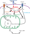
Glycolysis and glutaminolysis in osteoclast differentiation and bone-resorbing function. Due to the bioenergetic needs for osteoclastogenesis and bone resorpion, osteoclasts contain abundant mitochondria, the biogenesis of which is stimulated by PGC-1β according to our analysis (19). In addition, through the action of HIF-1α, the expression of Glut1 and glycolytic genes is stimulated toward the maturation stage, which contributes to the bone-resorbing function (20). The expression of Slc1a5 and glutaminase is induced via c-MYC early during the course of differentiation, which contributes to osteoclastogenesis and the bone-resorbing function (20). On net balance, osteoclast differentiation and function are positively and negatively regulated by mTOR and AMPK, respectively (20).
Fig. 4
Fig. 4
‘Clastokines’, the secreted products of osteoclasts, regulate osteogenesis and angiogenesis. As bone marrow macrophages (BMMs) become committed pre-osteoclasts (preOCs) and terminally differentiate into mature osteoclasts (mOCs), they produce and secrete various molecules, as illustrated in this figure. Different isoforms of PDGF are secreted at distinct differentiation stages (33), then binding to PDGFR-α on mesenchymal stromal cells (MSCs) or PDGFR-β on vascular cells, thereby regulating osteogenesis and angiogenesis. Along with differentiation, S1P production is increased, which acts on osteoblasts (OBs) through S1P3. The C3a that is locally processed from osteoclast-derived C3 stimulates osteoblastogenesis through C3aR (34). Finally, the mOCs engaged in bone resorption (active mOCs) produce particular molecules, such as Cthrc1 (collagen triple helix repeat containing 1), which favors osteoblast differentiation, diverting the cells from adipogenesis (35).

References

    1. Erlebacher A., Filvaroff E.H., Gitelman S.E., Derynck R. (1995) Toward a molecular understanding of skeletal development. Cell 80, 371–378 - PubMed
    1. Kronenberg H.M. (2003) Developmental regulation of the growth plate. Nature 423, 332–336 - PubMed
    1. Zaidi M. (2007) Skeletal remodeling in health and disease. Nat. Med. 13, 791–801 - PubMed
    1. Teitelbaum S.L. (2000) Bone resorption by osteoclasts. Science (New York, N.Y.) 289, 1504–1508 - PubMed
    1. Teitelbaum S.L., Ross F.P. (2003) Genetic regulation of osteoclast development and function. Nat. Rev. Genet. 4, 638–649 - PubMed

Publication types

MeSH terms

Substances