

Computed tomography diagnosis of a thoracic and abdominal penetrating foreign body in a dog
- PMID: 26538669
- PMCID: PMC4608467
Computed tomography diagnosis of a thoracic and abdominal penetrating foreign body in a dog
Abstract
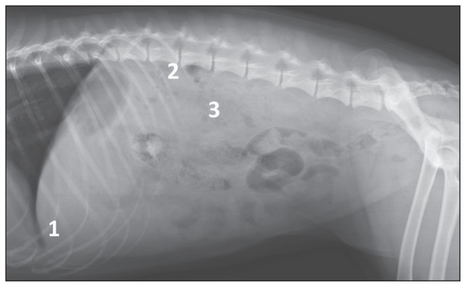
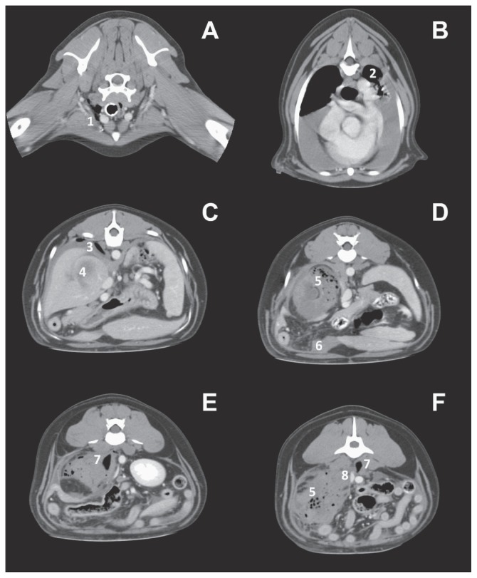
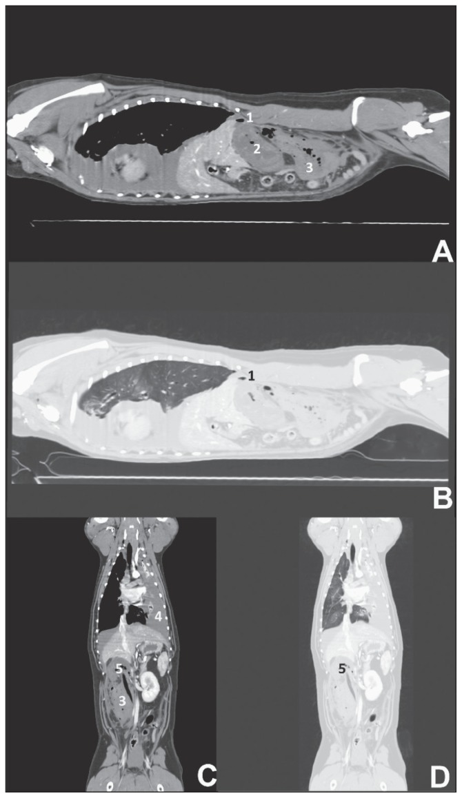
A 1.5-year-old, spayed female, mixed-breed dog was presented for hemoabdomen associated with an abdominal mass. Upon presentation bicavitary effusion was diagnosed. A penetrating intra-abdominal wooden foreign body was identified using computed tomography. This case describes a thoracic penetrating wooden foreign body causing bicavitary effusion following migration into the retroperitoneal space.
Diagnostic d’un corps étranger thoracique et abdominal pénétrant par tomodensitométrie chez un chien. Une chienne de race croisée stérilisée âgée de 1,5 ans a été présentée pour un hémoabdomen associé à une masse abdominale. Sur présentation, une effusion bicavitaire a été diagnostiquée. Un corps étranger en bois intra-abdominal pénétrant a été identifié par tomodensitométrie. Ce cas décrit un corps étranger en bois pénétrant dans le thorax et causant une effusion bicavitaire après la migration dans l’espace rétropéritonéal.(Traduit par Isabelle Vallières).
Figures
References
-
- White RAS, Lane JG. Pharyngeal stick penetration injuries in the dog. J Small Anim Pract. 1988;29:13–35.
-
- Laverty S, Lavoie JP, Pascoe JR, Ducharme N. Penetrating wounds of the thorax in 15 horses. Equine Vet J. 1996;28:220–224. - PubMed
-
- Girffiths LG, Tiruneh R, Sullivan M, Reid SWJ. Oropharyngeal penetrating injuries in 50 dogs: A retrospective study. Vet Surg. 2000;29:383–388. - PubMed
-
- Doran P, Wright CA, Moore AH. Acute oropharyngeal and esophageal stick injury in forty-one dogs. Vet Surg. 2008;37:781–785. - PubMed
-
- Halfacree NZ, Whatmough C, Mantis P, Baines S. Computed tomography as an aid to management of chronic oropharyngeal stick injury in the dog. J Small Anim Pract. 2008;49:451–457. - PubMed
Publication types
MeSH terms
LinkOut - more resources
Full Text Sources
Medical