. 2015 Oct;28(4):197-206.
doi: 10.1293/tox.2015-0020. Epub 2015 Jul 27.

Dietary curcumin supplementation attenuates 1-methyl-4-phenyl-1,2,3,6-tetrahydropyridine (MPTP) neurotoxicity in C57BL mice

Affiliations

Dietary curcumin supplementation attenuates 1-methyl-4-phenyl-1,2,3,6-tetrahydropyridine (MPTP) neurotoxicity in C57BL mice

Xi-Jun He et al. J Toxicol Pathol. 2015 Oct.

Erratum in

  • Errata (Printer's correction).
    [No authors listed] [No authors listed] J Toxicol Pathol. 2016 Jan;29(1):74. Epub 2016 Feb 17. J Toxicol Pathol. 2016. PMID: 26989306 Free PMC article.

Abstract

Studies in vivo and in vitro suggest that curcumin is a neuroprotective agent. Experiments were conducted to determine whether dietary supplementation with curcumin has neuroprotective effects in a mouse model of Parkinson's disease (PD). Treatment with 1-methyl-4-phenyl-1,2,3,6-tetrahydropyridine (MPTP) significantly induced the loss of dopaminergic cells in the substantia nigra and deletion of dopamine in the striatum, which was attenuated by long-term (7 weeks) dietary supplementation with curcumin at a concentration of 0.5% or 2.0% (w/w). Although curcumin did not prevent the MPTP-induced apoptosis of neuroblasts in the subventricular zone (SVZ), it promoted the regeneration of neuroblasts in the anterior part of the SVZ (SVZa) at 3 days after MPTP treatment. Furthermore, curcumin enhanced the MPTP-induced activation of microglia and astrocytes in the striatum and increased the expression of glial cell line-derived neurotrophic factor (GDNF) and transforming growth factor-β1 (TGFβ1) in the striatum and SVZ. GDNF and TGFβ1 are thought to play an important role in protecting neurons from injury in the central and peripheral nervous systems. These results suggest that long-term administration of curcumin blocks the neurotoxicity of MPTP in the nigrostriatal dopaminergic system of the mouse and that the neuroprotective effect might be correlated with the increased expression of GDNF and TGFβ1. Curcumin may be effective in preventing or slowing the progression of PD.

Keywords: MPTP; astrocyte; curcumin; dopamine; microglia; neuroprotective.


Figures

Fig. 1.
Fig. 1.
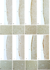
Effect of dietary curcumin supplementation on MPTP-induced destruction of the nigrostriatal dapaminergic system of mice shown by results of TH or DAT immunostaining. (A and F) Control group (basal diet + saline treatment). (B and G) Animals fed the basal diet and treated with MPTP showed a severe loss of dopaminergic neurons in the SNpc (B) and dopamine depletion in the striatum (G). (C and H) Animals fed a diet supplemented with 0.5% (w/w) curcumin for 7 weeks and then treated with MPTP. (D and I) Animals fed a diet supplemented with 2% (w/w) curcumin for 7 weeks and then treated with MPTP. (E) The number of TH-positive cells in the unilateral SNpc is presented as the mean ± SD. (J) The immunohistochemical staining intensity value for DAT in the unilateral striatum is presented as the mean ± SD; the value of saline control is set as 1.0. *p<0.05 versus the mice fed the basal diet and treated with MPTP; ** p<0.05 versus the mice fed the basal diet and treated with saline. Scale bar: A­–D, 100 μm; F–I, 500 μm.
Fig. 2.
Fig. 2.
Quantitative analysis of apoptotic cells by the TUNEL assay. There is no significant difference between the groups treated with MPTP and fed with or without curcumin. Data are shown as the mean ± SD.
Fig. 3.
Fig. 3.
Curcumin promotes the regeneration of neuroblasts in the SVZa post treatment with MPTP. Representative microphotographs of sections of the SVZa are stained with HE (A–D), Dcx (E–H), and PCNA (I–L) and double labled with Dcx (red) and PCNA (green) (M–P). (A, E, I and M) Control group (basal diet + saline). (B, F, J and N) Animals fed the basal diet and treated with MPTP showed a severe loss of neuroblasts in the SVZa. (C, G, K and O) Animals fed a diet supplemented with 0.5% (w/w) curcumin for 7 weeks and then treated with MPTP. (D, H, L and P) Animals fed a diet supplemented with 2% (w/w) curcumin for 7 weeks and then treated with MPTP. Arrows in M-P show representative Dcx and PCNA double-labeled cells. LV, lateral ventricle. Scale bar: 20 μm.
Fig. 4.
Fig. 4.
Quantitative analysis of cells in the bilateral SVZa with immunohistochemical staining by using an anti-Dcx or anti-PCNA antibody. The number of Dcx+ or PCNA+ cells is presented as the mean ± SD. *p<0.05 versus the mice fed the basal diet and treated with MPTP; ** p<0.05 versus the mice fed the basal diet and treated with saline.
Fig. 5.
Fig. 5.
Curcumin enhances the activation of astrocytes and microglia in the striatum post treatment with MPTP. Representative microphotographs of sections of the striatum are stained for GFAP (A–H) and Iba 1 (I–P). (A, E, I, and M) Control group (basal diet + saline). (B, F, J and N) Animals fed the basal diet and treated with MPTP showed increased expression of GFAP (B and F) and Iba 1 (I and N). (C, G, K and O) Animals fed a diet supplemented with 0.5% (w/w) curcumin for 7 weeks and then treated with MPTP. (D, H, L and P) Animals fed a diet supplemented with 2% (w/w) curcumin for 7 weeks and then treated with MPTP. (E–H) Higher magnification of A–D. (M–P) Higher magnification of I-L. Scale bar: A–D and I–L, 50 μm; E–H and M–P, 20 μm.
Fig. 6.
Fig. 6.
Quantitative analysis of cells in the unilateral striatum with immunohistochemical staining by using an anti-GFAP or anti-Iba antibody. The number of GFAP+ or Iba1+ cells is presented as the mean ± SD. * p<0.05 versus the mice fed the basal diet and treat with saline; **p<0.05 versus the mice fed the basal diet and treated with MPTP.
Fig. 7.
Fig. 7.
Curcumin increases the expression of GDNF and TGFβ-1 in the striatum and SVZ post treatment with MPTP. Representative microphotographs of sections of the SVZ (A–D, I–L) and striatum (E–H, M–P) stained for GDNF (A–H) or TGFβ-1 (I–P). (A, E, I and M) Control group (basal diet + saline). (B, F, J and N) Animals fed the basal diet and treated with MPTP. (C, G, K and O) Animals fed a diet supplemented with 0.5% (w/w) curcumin for 7 weeks and then treated with MPTP. (D, H, L and P) Animals fed a diet supplemented with 2% (w/w) curcumin for 7 weeks and then treated with MPTP. LV, lateral ventricle. Scale bar: A–D and I–L, 20 μm; E–H and M–P,100 μm.

References

    1. Langston JW, Langston EB, and Irwin I. MPTP-induced parkinsonism in human and non-human primates—clinical and experimental aspects. Acta Neurol Scand Suppl. 100: 49–54. 1984. - PubMed
    1. Chiueh CC, Markey SP, Burns RS, Johannessen JN, Jacobowitz DM, and Kopin IJ. Neurochemical and behavioral effects of 1-methyl-4-phenyl-1,2,3,6- tetrahydropyridine (MPTP) in rat, guinea pig, and monkey. Psychopharmacol Bull. 20: 548–553. 1984. - PubMed
    1. Heikkila RE, Cabbat FS, Manzino L, and Duvoisin RC. Effects of 1-methyl-4-phenyl-1,2,5,6-tetrahydropyridine on neostriatal dopamine in mice. Neuropharmacology. 23: 711–713. 1984. - PubMed
    1. Drechsel DA, and Patel M. Role of reactive oxygen species in the neurotoxicity of environmental agents implicated in Parkinson’s disease. Free Radic Biol Med. 44: 1873–1886. 2008. - PMC - PubMed
    1. Francis JW, Von Visger J, Markelonis GJ, and Oh TH. Neuroglial responses to the dopaminergic neurotoxicant 1-methyl-4-phenyl-1,2,3,6-tetrahydropyridine in mouse striatum. Neurotoxicol Teratol. 17: 7–12. 1995. - PubMed

LinkOut - more resources