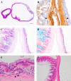
Vitelline cyst in the rat ileum
- PMID: 26538812
- PMCID: PMC4604132
- DOI: 10.1293/tox.JTP-2015-0021
Vitelline cyst in the rat ileum
Erratum in
-
Errata (Printer's correction).J Toxicol Pathol. 2016 Jan;29(1):74. Epub 2016 Feb 17. J Toxicol Pathol. 2016. PMID: 26989306 Free PMC article.
Abstract
Congenital vitelline duct anomalies other than Meckel's diverticulum are rare in animals. A cyst of approximately 8 mm in diameter was observed on the antimesenteric surface of the ileal serosa in a 10-week-old female Crl:CD(SD) rat. Microscopically, the cyst closely resembled the ileum, but it did not communicate with the ileal lumen. We diagnosed this case as a vitelline cyst derived from the vitelline duct based on the location where it developed and its histological behavior. In rats, only Meckel's diverticulum has been reported with a congenital anomaly of the vitelline duct, and no other spontaneous anomalies including a vitelline cyst have been reported. This case may be the first report concerning a vitelline cyst in the rat ileum.
Keywords: Meckel’s diverticulum; ileum; rat; vitelline cyst; vitelline duct.
Figures


References
-
- Bauer SB, and Retik AB. Urachal anomalies and related umbilical disorders. Urol Clin North Am. 5: 195–211. 1978. - PubMed
-
- Dencker L. Trypan blue accumulation in the embryonic gut of rats and mice during the teratogenic phase. Teratology. 15: 179–184. 1977. - PubMed
-
- Iwashita A. Intestine-Nonneoplastic disease. In: Surgical Pathology, 4th ed. Mukai K, Manabe T, and Fukayama M (eds). Bunkodo, Tokyo. 503–559. 2006. (in Japanese).
-
- DiSantis DJ, Siegel MJ, and Katz ME. Simplified approach to umbilical remnant abnormalities. Radiographics. 11: 59–66. 1991. - PubMed
-
- Levy AD, and Hobbs CM. From the archives of the AFIP. Meckel diverticulum: radiologic features with pathologic Correlation. Radiographics. 24: 565–587. 2004. - PubMed
Publication types
LinkOut - more resources
Full Text Sources
Other Literature Sources
