Myoepithelioma
- PMID: 26538968
- PMCID: PMC4606710
- DOI: 10.4103/0975-7406.163560
Myoepithelioma
Abstract
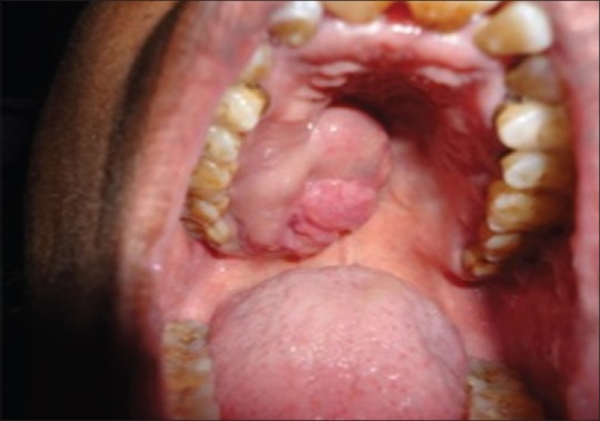
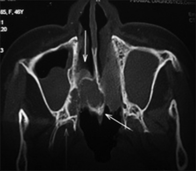
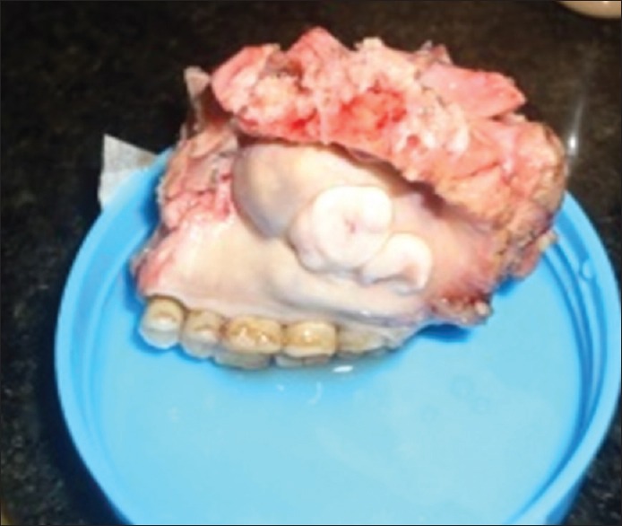
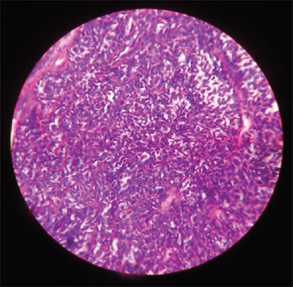
Myoepithelioma is a benign salivary gland tumor in the head and neck region, accounting for 1-1.5% of all glandular tumors. The diagnosis is rendered histopathologically, and it includes the proliferation of myoepithelial cells, without chondroid or myxochondroid stroma and ductal components (up to 5% of ductal component is acceptable). In our case report, this lesion has occurred in a 46-year-old female patient, and presented as well defined, nodular growth on the right posterior palatal region. Bony erosion and invasion were observed radiographically, and the lesion was excised surgically, with 1-2 cm of clear margin. The microscopic features included proliferating tumor sheets, composed of bland looking spindle and plasmacytoid shaped myoepithelial cells, and few cells showed clear cytoplasm, which were confirmed immunohistochemically as myoepithelial cells. Thus, the final diagnosis of benign myoepithelioma was rendered and no recurrence had been reported so far in the regular follow-up.
Keywords: Benign salivary gland lesion; myoepithelial cells; myoepithelioma.
Conflict of interest statement
Figures
References
-
- Barnes L, Appel BN, Perez H, El-Attar AM. Myoepithelioma of the head and neck: Case report and review. J Surg Oncol. 1985;28:21–8. - PubMed
-
- Kawashima Y, Kobayashi D, Ishikawa N, Kishimoto S. A case of myoepithelioma arising in an accessory parotid gland. J Laryngol Otol. 2002;116:474–6. - PubMed
-
- Sciubba JJ, Brannon RB. Myoepithelioma of salivary glands: Report of 23 cases. Cancer. 1982;49:562–72. - PubMed
-
- Morinière S, Robier A, Machet MC, Beutter P, Lescanne E. Massive infra-clinic invasion of the facial nerve by a myoepithelial carcinoma of the parotid. Int J Pediatr Otorhinolaryngol. 2003;67:663–7. - PubMed
-
- Sheldon WH. So-called mixed tumors of the salivary glands. Arch Pathol. 1943;35:1–20.
Publication types
LinkOut - more resources
Full Text Sources
Other Literature Sources