Review
. 2015 Oct 21:9:419.
doi: 10.3389/fncel.2015.00419. eCollection 2015.

Ion dynamics during seizures

Affiliations
Review

Ion dynamics during seizures

Joseph V Raimondo et al. Front Cell Neurosci. .

Abstract

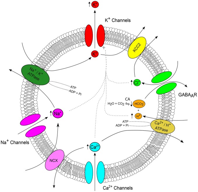
Changes in membrane voltage brought about by ion fluxes through voltage and transmitter-gated channels represent the basis of neural activity. As such, electrochemical gradients across the membrane determine the direction and driving force for the flow of ions and are therefore crucial in setting the properties of synaptic transmission and signal propagation. Ion concentration gradients are established by a variety of mechanisms, including specialized transporter proteins. However, transmembrane gradients can be affected by ionic fluxes through channels during periods of elevated neural activity, which in turn are predicted to influence the properties of on-going synaptic transmission. Such activity-induced changes to ion concentration gradients are a feature of both physiological and pathological neural processes. An epileptic seizure is an example of severely perturbed neural activity, which is accompanied by pronounced changes in intracellular and extracellular ion concentrations. Appreciating the factors that contribute to these ion dynamics is critical if we are to understand how a seizure event evolves and is sustained and terminated by neural tissue. Indeed, this issue is of significant clinical importance as status epilepticus-a type of seizure that does not stop of its own accord-is a life-threatening medical emergency. In this review we explore how the transmembrane concentration gradient of the six major ions (K(+), Na(+), Cl(-), Ca(2+), H(+)and [Formula: see text]) is altered during an epileptic seizure. We will first examine each ion individually, before describing how multiple interacting mechanisms between ions might contribute to concentration changes and whether these act to prolong or terminate epileptic activity. In doing so, we will consider how the availability of experimental techniques has both advanced and restricted our ability to study these phenomena.

Keywords: calcium; chloride; epilepsy; ion dynamics; pH; potassium; seizures; sodium.


Figures

Figure 1
Figure 1
Ion concentration changes during seizures. Top, an intracellular recording from a CA3 hippocampal neuron during a seizure-like event in vitro demonstrates typical neuronal membrane potential changes which accompany seizure episodes. Seizures consist of a “preictal” phase, a “tonic” phase (characterized by membrane potential depolarization upon which occur high-frequency, low-amplitude discharges, dark gray), a “clonic” or “after-discharge phase” (composed of rhythmic bursts of activity that emerge from a relatively hyperpolarized membrane potential background, light gray) and lastly a “postictal” phase (characterized by a hyperpolarized membrane potential lasting for several minutes before slowly recovering to preseizure values). Bottom, representative changes in ion concentration associated with seizures. The accompanying traces should be viewed as “best approximations” based on the available literature. [K+]e is thought to peak at the end of the tonic phase with an undershoot accompanying the postictal period (Dreier and Heinemann, ; Fröhlich et al., 2008). The [Na+]i trace is inferred from experimental measurements and supported by computational modeling work, which suggests that [Na+]i is likely to peak at the end of the seizure (Krishnan and Bazhenov, 2011). [Ca2+]e has been shown to drop rapidly to approximately 100 μM during an ictal event (Pumain et al., 1985). [Cl]i increases during seizures and is highest during the clonic phase (Raimondo et al., ; Ellender et al., 2014). Neuronal pHi decreases during the seizure, reaching its minimum around the time of seizure offset, and then slowly recovering during the postictal phase (Xiong et al., ; Raimondo et al., 2012a).
Figure 2
Figure 2
Ion interactions during seizures. The major ion channels and transporters, which serve as important nodes of interaction between ions are depicted, with arrows highlighting the typical direction of ionic flux. Arrows adjacent to an ion reflect the direction of seizure induced concentration change (see Figure 1). During a seizure, K+ efflux via a host of K+ channels (red) results in accumulation of extracellular K+. The cation-chloride transporter KCC2 (yellow) serves as an important link between the seizure-associated reduction in the transmembrane K+ gradient and intracellular Cl accumulation via GABAA receptors (GABAARs) (green). GABAARs, which are permeable to both Cl and HCO3 connect the regulation of Cl, HCO3 and pH via carbonic anhydrases which catalyze the reversible reaction of H2O and CO2 to HCO3 and H+ (orange). Seizures are associated with intracellular acidification which is due, in part, to the activity of Ca2+/H+ ATPase as it imports H+ and extrudes Ca2+ in attempt to restore baseline Ca2+ concentrations following activity-induced Ca2+ influx (cyan). Na+/Ca2+ exchangers (NCX, pink) connect Ca2+ and Na+ concentration. Seizure-associated Na+ influx via voltage and ligand gated Na+ channels (magenta) up regulates the activity of Na+/K+ ATPases (forest green). Finally, increased intra-neuronal concentrations of Cl, H+, Ca2+ and Na+ all activate K+ channels (dashed gray line). The number and complexity of possible ionic interactions highlights the importance of computational models for determining the relevance of these continuously evolving variables, which are often difficult to study experimentally.

References

    1. Abad M. F. C., Di Benedetto G., Magalhães P. J., Filippin L., Pozzan T. (2004). Mitochondrial pH monitored by a new engineered green fluorescent protein mutant. J. Biol. Chem 279, 11521–11529. 10.1074/jbc.m306766200 - DOI - PubMed
    1. Amzica F., Steriade M. (2000). Neuronal and glial membrane potentials during sleep and paroxysmal oscillations in the neocortex. J. Neurosci. 20, 6648–6665. - PMC - PubMed
    1. Arosio D., Ricci F., Marchetti L., Gualdani R., Albertazzi L., Beltram F. (2010). Simultaneous intracellular chloride and pH measurements using a GFP-based sensor. Nat. Methods 7, 516–518. 10.1038/nmeth.1471 - DOI - PubMed
    1. Avoli M., Louvel J., Pumain R., Olivier A. (1987). Seizure-like discharges induced by lowering [Mg2+]o in the human epileptogenic neocortex maintained in vitro. Brain Res. 417, 199–203. - PubMed
    1. Balestrino M., Somjen G. G. (1988). Concentration of carbon dioxide, interstitial pH and synaptic transmission in hippocampal formation of the rat. J. Physiol. 396, 247–266. 10.1113/jphysiol.1988.sp016961 - DOI - PMC - PubMed