. 2015 Nov 4;88(3):499-513.
doi: 10.1016/j.neuron.2015.09.048.

Genes that Affect Brain Structure and Function Identified by Rare Variant Analyses of Mendelian Neurologic Disease

Affiliations

Genes that Affect Brain Structure and Function Identified by Rare Variant Analyses of Mendelian Neurologic Disease

Ender Karaca et al. Neuron. .

Abstract

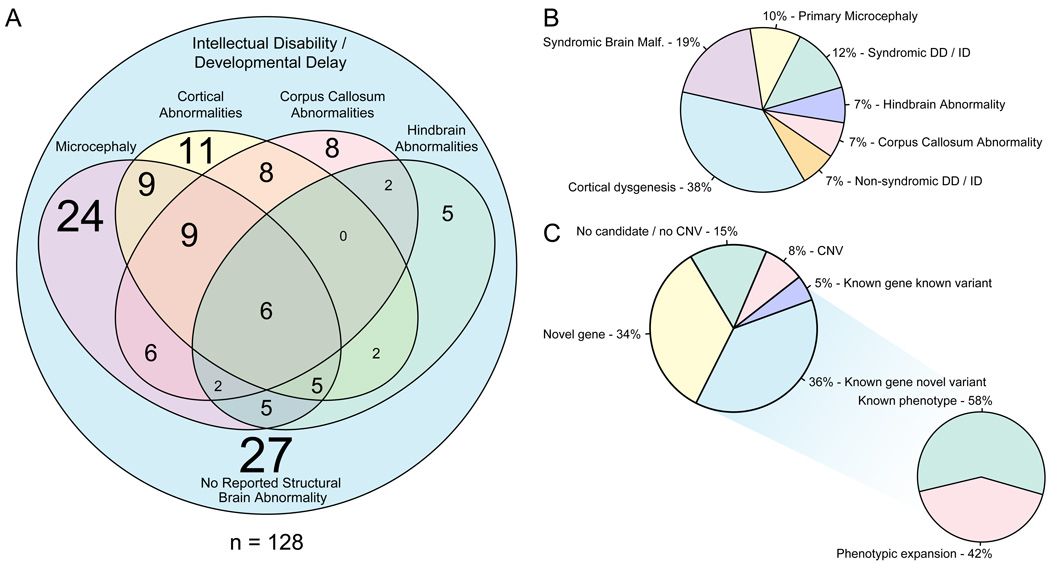
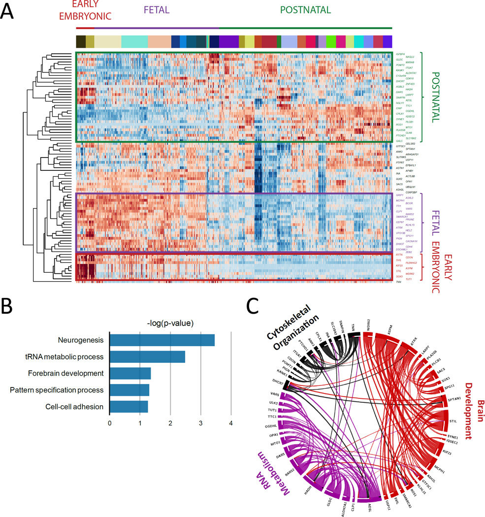
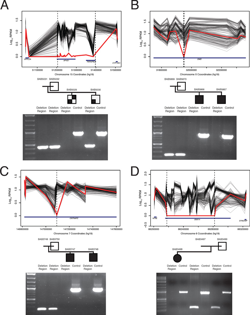
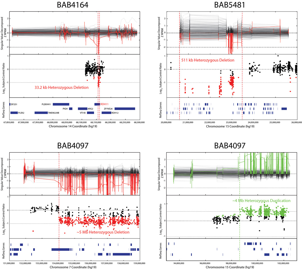
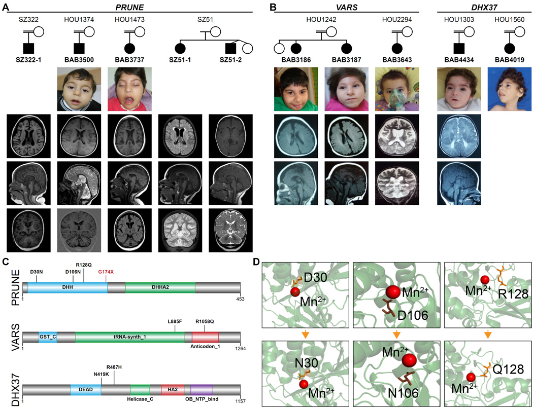
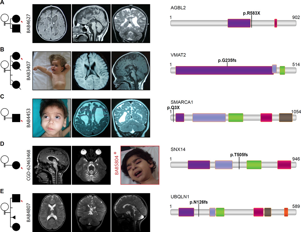
Development of the human nervous system involves complex interactions among fundamental cellular processes and requires a multitude of genes, many of which remain to be associated with human disease. We applied whole exome sequencing to 128 mostly consanguineous families with neurogenetic disorders that often included brain malformations. Rare variant analyses for both single nucleotide variant (SNV) and copy number variant (CNV) alleles allowed for identification of 45 novel variants in 43 known disease genes, 41 candidate genes, and CNVs in 10 families, with an overall potential molecular cause identified in >85% of families studied. Among the candidate genes identified, we found PRUNE, VARS, and DHX37 in multiple families and homozygous loss-of-function variants in AGBL2, SLC18A2, SMARCA1, UBQLN1, and CPLX1. Neuroimaging and in silico analysis of functional and expression proximity between candidate and known disease genes allowed for further understanding of genetic networks underlying specific types of brain malformations.


Figures

FIGURE 1
FIGURE 1. Phenotypic clustering of the cohort and summary of WES findings
A. Venn diagram of clinical and neuro-radiological features. The font size of the numbers correlates with the number of individuals that represent any given category. B. Phenotypic clustering of the probands according to their most outstanding feature revealed seven major groups: primary microcephaly (10%), cortical dysgenesis (38%), callosal abnormalities (7%), hindbrain malformations (7%), syndromic brain malformations (19%), nonsyndromic intellectual disability (7%), and syndromic developmental delay or intellectual disability (12%). C. WES analysis revealed novel candidates in 34%, novel variants in known genes in 36%, known variants in known genes in 5%, CNVs in 8% of the families. 42% of the families with novel variants in known genes represent phenotypic expansion.
FIGURE 2
FIGURE 2. Expression, annotation, and pathway analysis of known and candidate genes
A. Unsupervised clustering based on mRNA levels in the brain tissue partitioned the known and candidate genes into 4 subgroups: genes expressed only in early embryonic development, only in fetal development, only in adult brain tissue, and lastly in both embryonic development and adult tissue. KIF23, TUT1, CLP1, PRUNE, VARS, and DHX37 are included among the genes expressed only in early embryonic development or in fetal development. B. Biological functional annotation of the novel and known mutated genes in our cohort revealed that they were most significantly enriched in neurogenesis, tRNA metabolic process, forebrain development, pattern specification process, and cell-cell adhesion. C. The protein-protein interaction network had a greater degree of connectivity than expected by chance (P-value=3.26*10-3). This network revealed 3 highly interconnected protein networks, consisting of genes significantly enriched in brain development, RNA metabolism, and cytoskeletal organization.
FIGURE 3
FIGURE 3. Homozygous and hemizygous CNV’s
A. Homozgous deletion encompassing AP4E1 in BAB5029 but not BAB5030. B. Hemizygous intragenic deletion of DMD interrupting exons 46 and 47; C. Homozygous intragenic deletion of CNTNAP2; and D. Homozygous deletion almost entirely encompassing SNX14. PCR validation underneath each pedigree shows amplification or lack thereof of the deletion region and a positive control PCR of an unrelated locus. Amplification of the deletion region in parents and unaffected siblings indicates either a heterozygous (assumed for parents, as obligate carriers) or homozygous wild type state.
FIGURE 4
FIGURE 4. Heterozygous CNVs identified by whole exome sequencing
Upper panel represents CNV as predicted from WES data; middle panel represents validation by array studies; and lower panel shows chromosomal position and RefSeq genes involved. Parental studies for CNVs were beyond the scope of this article.
FIGURE 5
FIGURE 5. Patients with mutations in PRUNE VARS and DHX37
A. Pedigrees of the families with PRUNE mutations show that 3 families (BAB3500 and BAB3737 are of Turkish origin, SZ322 is of Saudi origin) are consanguineous while SZ51 (US origin) is not. Available patient images reveal some dysmorphic features most probably a result of microcephaly. Note that axial, mid-sagittal and coronal slices from the brain MRIs of each patient demonstrate a similar phenotype consisting of cortical atrophy, thin/hypoplastic corpus callosum and prominent cerebellar atrophy. B. Families with homozygous VARS and DHX37 mutations presented with severe microcephaly, DD/ID, and cortical atrophy. C. The human PRUNE is a member of DHH superfamily, and contains DHH and DHHA protein domains at the N-l and C-termini respectively. Also note that human VARS is a multi-domain protein, containing N-terminal glutathione S-transferase (GST_N), C-terminal glutathione S-transferase (GST_C), tRNA synthase class I (tRNA-synth_1) and anticodon-binding domain of tRNA (anticodon_1). L885F and R1058Q substitutions occur in the latter two domains respectively. DHX37 protein contains DEAD, Helicase_C, HA2 and OB_NTP_binding domains. N419K substitution occurs nearby the DEAD domain, which plays a role in several aspects of RNA metabolism processes such as translation initiation and pre-mRNA splicing, whereas the other substitution (p.R487H) locates between DEAD and Helicase_C domains. D. DHH domain of PRUNE carries a highly conserved motif of Asp-His-His (DHH). The aspartic acid in DHH motif of the human PRUNE was shown to bind Mg2+ (D’Angelo et al., 2004). Model structure for human PRUNE protein from SwissModel repository suggests that negatively charged D30 and D106 interact directly with the positively charged cofactor, while R128 and G174 are in close proximity to the catalytic site.
FIGURE 6
FIGURE 6. Pedigrees, clinical and radiologic images of patients with homozygous LOF mutations
Consanguinity between parents is indicated in each pedigree. A. Brain MRI of BAB4627 revealed severe cortical dysplasia, diffuse hypoplastic corpus callosum, dilated lateral ventricles, simplified gyral pattern, and dysmorphic basal ganglia. Note the similarity of the brain phenotype in BAB4627, with homozygous AGBL2 p. R583X variant, to tubulinopathy-related cortical dysplasia syndromes. B. BAB3407’s MRI presents cortical atrophy, thin and dysplastic corpus callosum. Patient image illustrates her severe dystonia. C. BAB4453 with homozygous stop gain (p.Q3X) in SMARCA1 represents severe cortical atrophy. Patient image underlines the coarse face, bushy eyebrows, facial hypertrichosis, and long eye lashes which resembles the facial dysmorphism in Coffin-Siris syndrome. D. A homozygous frameshift mutation (p.505fs) was detected in SNX14 in patient CGD-62463468; note the MRI shows severe cerebellar atrophy. For comparison an image of a patient (BAB5804) from a different family with homozygous SNX14:c.T2390G:p.L797R mutation is provided and which also revealed a coarse face in the patient. E. The MRI of BAB4807 with homozygous p.N126fs in UBQLN1 shows the dolichocephalic appearance of the head, dilated lateral ventricles and Arnold-Chiari malformation.
FIGURE 7
FIGURE 7. Suggested correlation between neurodevelopmental stage, representative process, strong candidate genes, and phenotype
Selected genes and their protein-protein interactions are shown in terms of correlation with neurodevelopmental process and resultant phenotype.

References

    1. Akizu N, Cantagrel V, Zaki MS, Al-Gazali L, Wang X, Rosti RO, Dikoglu E, Gelot AB, Rosti B, Vaux KK, et al. Biallelic mutations in SNX14 cause a syndromic form of cerebellar atrophy and lysosome-autophagosome dysfunction. Nature genetics. 2015;47:528–534. - PMC - PubMed
    1. Alazami AM, Patel N, Shamseldin HE, Anazi S, Al-Dosari MS, Alzahrani F, Hijazi H, Alshammari M, Aldahmesh MA, Salih MA, et al. Accelerating novel candidate gene discovery in neurogenetic disorders via whole-exome sequencing of prescreened multiplex consanguineous families. Cell reports. 2015;10:148–161. - PubMed
    1. Alsiary R, Bruning-Richardson A, Bond J, Morrison EE, Wilkinson N, Bell SM. Deregulation of microcephalin and ASPM expression are correlated with epithelial ovarian cancer progression. PloS one. 2014;9:e97059. - PMC - PubMed
    1. Aravind L, Koonin EV. A novel family of predicted phosphoesterases includes Drosophila prune protein and bacterial RecJ exonuclease. Trends in biochemical sciences. 1998;23:17–19. - PubMed
    1. Bainbridge MN, Hu H, Muzny DM, Musante L, Lupski JR, Graham BH, Chen W, Gripp KW, Jenny K, Wienker TF, et al. De novo truncating mutations in ASXL3 are associated with a novel clinical phenotype with similarities to Bohring-Opitz syndrome. Genome medicine. 2013;5:11. - PMC - PubMed

Publication types

Associated data