. 2015 Nov 5;6(11):e1965.
doi: 10.1038/cddis.2015.315.

L-Stepholidine rescues memory deficit and synaptic plasticity in models of Alzheimer's disease via activating dopamine D1 receptor/PKA signaling pathway

Affiliations

L-Stepholidine rescues memory deficit and synaptic plasticity in models of Alzheimer's disease via activating dopamine D1 receptor/PKA signaling pathway

J-R Hao et al. Cell Death Dis. .

Abstract

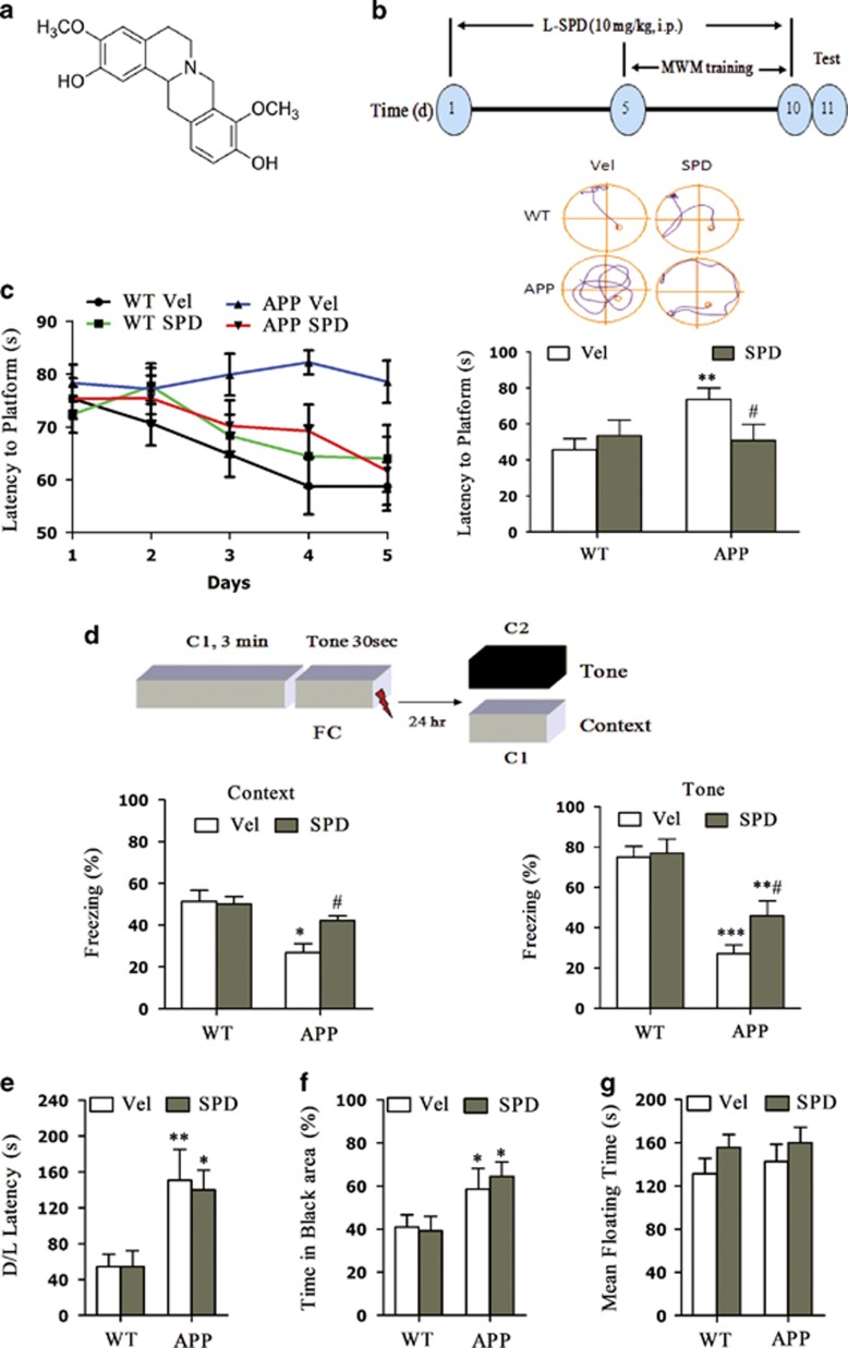
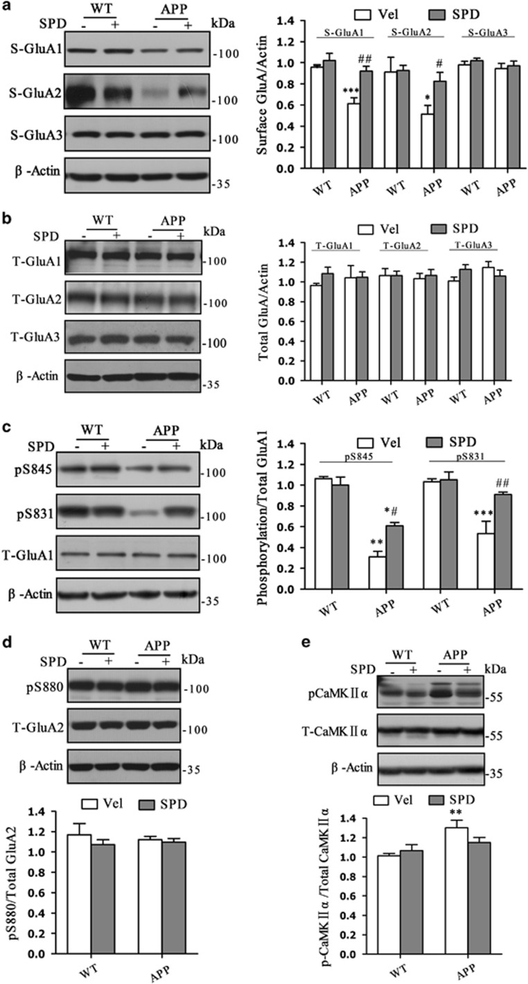
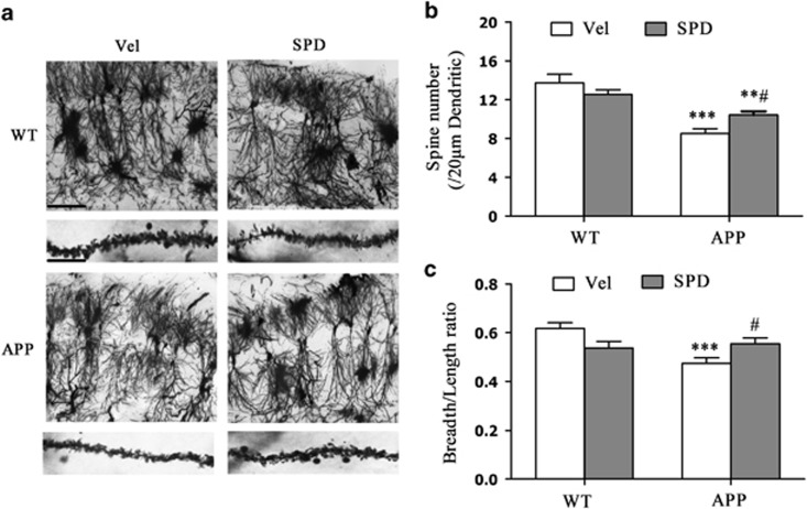
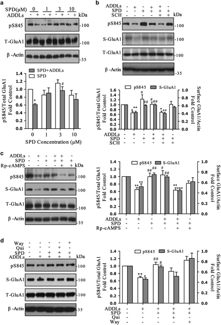
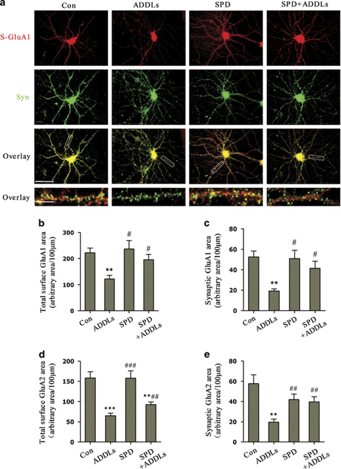
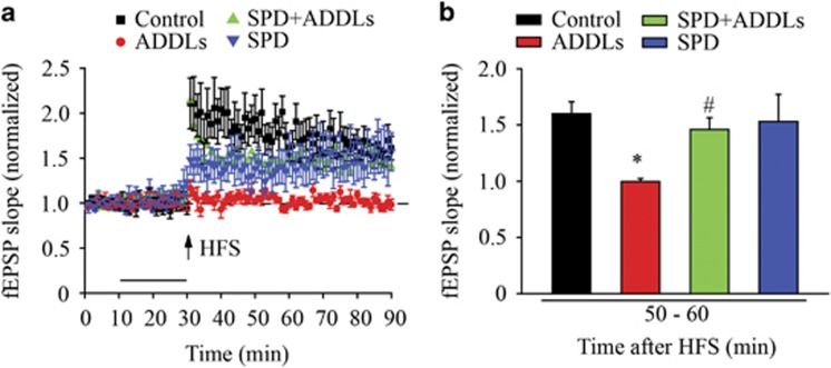
It is accepted that amyloid β-derived diffusible ligands (ADDLs) have a prominent role in triggering the early cognitive deficits that constitute Alzheimer's disease (AD). However, there is still no effective treatment for preventing or reversing the progression of the disease. Targeting α-amino-3-hydroxy-5-methylisoxazole-4-propionic acid (AMPA) receptor trafficking and its regulation is a new strategy for AD early treatment. Here we investigate the effect and mechanism of L-Stepholidine (L-SPD), which elicits dopamine D1-type receptor agonistic activity, while acting as D2-type receptor antagonist on cognition and synaptic plasticity in amyloid precursor protein (APP) and presenilin 1 (PS1) double-transgenic (APP/PS1) mice, and hippocampal cultures or slices treated with ADDLs. L-SPD could improve the hippocampus-dependent memory, surface expression of glutamate receptor A (GluA1)-containing AMPA receptors and spine density in hippocampus of APP/PS1 transgenic mice. L-SPD not only rescued decreased phosphorylation and surface expression of GluA1 in hippocampal cultures but also protected the long-term potentiation in hippocampal slices induced by ADDLs. Protein kinase A (PKA) agonist Sp-cAMPS or D1-type receptor agonist SKF81297 had similar effects, whereas PKA antagonist Rp-cAMPS or D1-type receptor antagonist SCH23390 abolished the effect of L-SPD on GluA1 trafficking. This was mediated mainly by PKA, which could phosphorylate serine residue at 845 of the GluA1. L-SPD may be explored as a potential therapeutic drug for AD through a mechanism that improves AMPA receptor trafficking and synaptic plasticity via activating D1/PKA signaling pathway.


Figures

Figure 1
Figure 1
L-SPD improves impaired learning and memory in APP/PS1 mice. (a) Chemical structure of L-SPD. (b) Schedule of test. (c) APP/PS1 mice (APP) injected with L-SPD (10 mg/kg, i.p.) spent less time on reaching the platform than that of DMSO vehicle (Veh) after training (n=16 in each group). The latency to first reach the platform was recorded 24 h after last training session. Representative routes to reach the platform are on the top. APP mice injected with vehicle spent more time on reaching the platform compared with WT mice injected with vehicle, whereas mice injected with L-SPD spent less time than that of the vehicle (n=11 in WT/Vel group, n=10 in WT/SPD group, n=14 in APP/Vel group, n=9 in APP/SPD group). (d) L-SPD significantly improved the impaired context-dependent fear memory and partly improved the impaired tone-dependent fear memory in APP/PS1 mice compared with WT mice (n=8 in each group). (e and f) L-SPD did not affect anxiety-like behavior. D/L test showed increased latency to exit the dark area (e) and the percentage of time spent in dark area (f) in APP/PS1 mice compared with WT mice treated with vehicle. L-SPD did not affect the latency and time spent on black area (n=7 in APP/Vel group, n=8 in other groups). (g) L-SPD did not affect depression-like behavior. Forced swim test showing similar time spent on floating in APP/PS1 mice compared with WT mice treated with vehicle. L-SPD did not affect the mean floating time (n=13 in APP/Vel group, n=15 in other groups). *P<0.05, **P<0.01, ***P<0.001 versus corresponding WT group; #P<0.05 versus APP/vehicle group. Data are represented as mean±S.E.M
Figure 2
Figure 2
L-SPD rescues the decreased phosphorylation and surface expression of GluA1-containing AMPA receptors in the hippocampus of APP/PS1 mice. (a) Surface expression of GluA1 and GluA2 subunits were decreased, whereas S-GluA3 expression was not changed in APP/PS1 mice treated with vehicle. L-SPD significantly improved the levels of surface expression of GluA1 and GluA2 in APP/PS1 mice. (GluA1: n=4 in each group, GluA2: n=5 in each group, GluA3: n=4 in each group). (b) Total expression of GluA1, GluA2 and GluA3 subunits were not changed (n=4 in each group). (c) Phosphorylated levels of both pS845 and pS831 of GluA1 were decreased in APP/PS1 mice treated with vehicle. L-SPD could improve the levels of both pS845 and pS831 (n=4 in each group). (d) Phosphorylated levels of pS880 of GluA2 was not changed and L-SPD had no effect on pS880 in APP/PS1 mice (n=3 in each group). (e) Phosphorylated levels of CaMKIIα was increased in APP /PS1 mice treated with vehicle. L-SPD had no effect on p-CaMKIIα (n=6 in each group). *P<0.05, **P<0.01, ***P<0.001 versus corresponding WT group; #P<0.05, ##P<0.01 versus APP/vehicle group. Data are represented as mean±S.E.M
Figure 3
Figure 3
L-SPD improves spine density in the hippocampus of APP/PS1 mice. (a) Golgi staining of the hippocampal CA1 area (n=6 in each group). Scale bars= 70 μm (top panel); 15 μm (bottom panel). (b) Quantitative analysis of spine density. The spine number was decreased in the APP/PS1 mice treated with vehicle. L-SPD partly rescued the impaired spine density (n=20 in each group). (c) Quantitative analysis of breadth-to-length ratio. The ratio was decreased in the APP/PS1 mice treated with vehicle. L-SPD significantly improved the impaired ratio (n=51 in each group). **P<0.01, ***P<0.001 versus corresponding WT group; #P<0.05 versus APP/vehicle group. Data are represented as mean±S.E.M
Figure 4
Figure 4
Activation of D1/PKA rescues the decreased GluA1-containing AMPA receptor surface expression induced by ADDLs in cultured hippocampal neurons. (ac) Surface expression of GluA1 (S-GluA1) and GluA2 (S-GluA2) were significantly decreased after treating with ADDLs (500 nM) for 3–24 h, whereas surface expression of GluA3 (S-GluA3) was not changed. Total expression of GluA1-3 (T-GluA) were not changed. (GluA1: n=5 in each group, GluA2: n=4 in each group, GluA3: n=4 in each group). (d) Phosphorylated levels of GluA1 at Serine 845 (pS845) and Serine 831 (pS831) were decreased after treating with ADDLs (pS845: n=9 in each group, pS831: n=9 in each group). (e) Pretreatment with PKA activator Sp-cAMPS (10 μM) rescued the decreased pS845 and surface expression of GluA1 induced by ADDLs, whereas the PKA inhibitor Rp-cAMPS (10 μM) reversed these effects. Sp-cAMPS or Rp-cAMPS was added 10 min before ADDLs (n=4 in each group). (f) Pretreatment with D1-type receptor agonist SKF 81297 (SKF, 3 μM) rescued the decreased pS845 and surface expression of GluA1 induced by ADDLs, whereas the antagonist SCH 23390 (SCH, 10 μM) reversed these effects. SKF 81297 or SCH 23390 was added 15 min before ADDLs (n=6 in each group). *P<0.05, **P<0.01, ***P<0.001 versus corresponding control group (Con); #P<0.05, ##P<0.01, ###P<0.001 versus corresponding ADDL group. Data are represented as mean±S.E.M
Figure 5
Figure 5
L-SPD prevents ADDL-induced loss of surface expression of GluA1 through activation of D1/PKA signal pathway in cultured hippocampal neurons. (a) Pretreatment with L-SPD (3 μM, 15 min) rescued pS845 of GluA1, for which phosphorylation was decreased induced by ADDLs. There was a trend toward increased pS845 under higher (10 μM) or lower (1 μM) concentration of L-SPD (n=8 in each group). *P<0.05 versus control group without L-SPD; #P<0.05 versus ADDLs group without L-SPD (n=4 in each group). (b) L-SPD (3 μM) rescued both the decreased pS845 and surface expression of GluA1 induced by ADDLs, whereas SCH 23390 (SCH, 10 μM) abolished these effects (n=4 in each group). (c) Rp-cAMPS (10 μM) abolished the effects of L-SPD (3 μM) on pS845 (n=5 in each group) and surface expression of GluA1 (n=4 in each group). (d) Co-administration L-SPD (3 μM) with D2 receptor agonist Quinpirole hydrochloride (Qui, 1 μM) or 5-HTA receptor antagonist WAY-100635 maleate salt (Way, 1 μM) had no significant effect on the role of L-SPD in S-GluA1 expression and pS845 level (n=4 in each group). *P<0.05, **P<0.01, ***P<0.001 versus corresponding control group; #P<0.05, ##P<0.01 versus corresponding ADDL group. Data are represented as mean±S.E.M
Figure 6
Figure 6
L-SPD rescues the decreased externalization and synaptic expression of GluA1/GluA2 in cultured hippocampal neurons induced by ADDLs. (a) Double staining against N-terminal S-GluA1 (S-GluA1, red) and synaptophysin (Syn, green) showed that pretreatment with L-SPD (3 μM) rescued both the decreased total surface and synaptic (yellow) expression of GluA1 induced by ADDLs (500 nM, 3 h). No change in synaptophysin was observed. Scale bars=100 μm (top three panels); 15 μm (bottom panel). (b) Quantitative analysis of total S-GluA1 area. (c) Quantitative analysis of synaptic GluA1 area. Error bars indicate S.E.M. from at least three independent experiments with 25 cells imaged per experimental condition in each experiment. (d) Quantitative analysis of total S-GluA2 area. (e) Quantitative analysis of synaptic GluA2 area. Error bars indicate S.E.M. from at least three independent experiments with 27 cells imaged per experimental condition in each experiment. **P<0.01, ***P<0.001 versus control group (Con); #P<0.05, ##P<0.01, ###P<0.001 versus ADDLs group
Figure 7
Figure 7
L-SPD rescues LTP impairment in hippocampal slices induced by ADDLs. (a) Delivery of high-frequency stimulation (HFS, 100 Hz) induced LTP in the CA1 of the hippocampus. Exposure of ADDLs (500 nM) inhibited LTP induction. Pre-incubation of L-SPD (3 μM) significantly improved the inhibition of LTP induced by ADDLs. L-SPD itself could also induce LTP under HFS. (b) Analysis of LTP responded at 60 min after HFS (n=6 in each group). *P<0.05 versus control group (Con); #P<0.05 versus ADDLs group. Data are represented as mean±S.E.M

Similar articles

Cited by

References

    1. 1Krafft GA, Klein WL. ADDLs and the signaling web that leads to Alzheimer's disease. Neuropharmacology 2010; 59: 230–242. - PubMed
    1. 2Ferreira ST, Klein WL. The Aβ oligomer hypothesis for synapse failure and memory loss in Alzheimer's disease. Neurobiol Learn Mem 2011; 96: 529–543. - PMC - PubMed
    1. 3Malinow R, Malenka RC. AMPA receptor trafficking and synaptic plasticity. Annu Rev Neurosci 2002; 25: 103–126. - PubMed
    1. 4Ren SQ, Yan JZ, Zhang XY, Bu YF, Pan WW, Yao W et al. PKCλ is critical in AMPA receptor phosphorylation and synaptic incorporation during LTP. EMBO J 2013; 32: 1365–1368. - PMC - PubMed
    1. 5Whitehead G, Jo J, Hogg EL, Piers T, Kim DH, Seaton G et al. Acute stress causes rapid synaptic insertion of Ca2+ -permeable AMPA receptors to facilitate long-term potentiation in the hippocampus. Brain 2013; 136: 3753–3765. - PMC - PubMed

Publication types

MeSH terms